Epidemiology
Etiology
- HIV-1
- Epidemiology: Cause of the vast majority (>95%) of HIV infections globally. Responsible for the AIDS pandemic.
- Virulence: More virulent; faster progression to AIDS and higher viral loads.
- Transmission: More easily transmitted than HIV-2.
- HIV-2
- Epidemiology: Primarily concentrated in West Africa.
- Virulence: Less virulent; slower disease progression and lower viral loads.
- Transmission: Less efficiently transmitted.
- Treatment Consideration: Intrinsically resistant to Non-Nucleoside Reverse Transcriptase Inhibitors (NNRTIs).
- Viral Tropism (Coreceptor Usage for Entry)
- To enter a host cell, HIV must bind to CD4 and a coreceptor. The coreceptor used determines the tropism.
- R5-Tropic (Macrophage-tropic)
- Uses the CCR5 coreceptor.
- Found on macrophages and T-helper cells.
- This is the dominant viral type during initial infection and the asymptomatic phase.
- Targeted by the drug Maraviroc, a CCR5 antagonist. A tropism assay is required before starting this medication.
- X4-Tropic (T-cell-tropic)
- Uses the CXCR4 coreceptor.
- Found primarily on T-helper cells.
- This variant tends to emerge in later stages of the disease.
- Appearance of X4-tropic virus is associated with a more rapid decline in CD4+ cells and faster progression to AIDS.
Pathophysiology
- Key Genes:
env: Glycoproteins gp120 (for host CD4 receptor binding) & gp41 (for fusion and entry).gag: Capsid protein p24.pol: Reverse transcriptase, integrase, protease.- HIV pol gene mutations are responsible for acquired resistance to treatment. Mutations of the env gene enable escape from host-neutralizing antibodies.
Natural history of HIV infection
- HIV infects CD4+ T-helper cells, macrophages, and dendritic cells.
- gp120 binds to CD4 on T-cell.
- gp120 then binds to a coreceptor: CCR5 (early infection, macrophages) or CXCR4 (late infection, T-cells).
- gp41 mediates fusion with the host cell membrane.
- Viral RNA is reverse transcribed into DNA by reverse transcriptase.
- Viral DNA is integrated into the host genome by integrase.
- Host machinery is used to produce viral proteins, which are cleaved by protease to form mature virions.
Clinical features
Acute HIV infection
- Timing: Develops 2–4 weeks post-exposure (occurs in ~50–90% of newly infected patients).
- Mononucleosis-like Syndrome:
- Fever, fatigue, generalized malaise, myalgias, arthralgias.
- Generalized non-tender lymphadenopathy.
- Pharyngitis and tonsillar swelling without exudates.
- Mucocutaneous Findings:
- Painful mucocutaneous ulcers (oral cavity, palate, esophagus, or genitalia) – highly specific for acute HIV vs EBV/CMV.
- Erythematous maculopapular rash (primarily trunk and face, non-pruritic).
- Neurological & GI Symptoms:
- Aseptic meningitis (headache, photophobia), peripheral neuropathy, facial palsy. c
- Nausea, vomiting, diarrhea, weight loss.
Tip
Acute retroviral syndrome is associated with extremely high levels of viral replication (~5 million copies/mL) as the cell-mediated and humoral antibody response against the virus is not yet fully activated. Therefore, laboratory results during this period usually show evidence of HIV in the plasma (positive viral load and p24 antigen) with a negative serologic response (negative HIV-1/HIV-2 antibody). This is referred to as the “window period,” as patients are infected with HIV but HIV antibody screening tests may be negative (newer screening tests incorporate testing for HIV p24 antigen and are more sensitive in early infection).
HIV-associated conditions
AIDS-defining conditions
CD4+ cell count < 500/mm3
- Kaposi sarcoma (typically occurs at CD4 count < 500)
- Lymphoma
- Mycobacterium tuberculosis
CD4+ cell count < 200/mm3
- Pneumocystis pneumonia
- Disseminated or extrapulmonary histoplasmosis
- Progressive multifocal leukoencephalopathy
CD4+ cell count < 100/mm3
- Cerebral toxoplasmosis
- Extrapulmonary cryptococcosis (especially cryptococcal meningitis)
- Cryptosporidiosis
- Etiology: Cryptosporidium species
- Clinical features: chronic, watery diarrhea (lasting > 1 month) with nausea and abdominal pains; typically at CD4 counts < 100
- Diagnostics: acid-fast oocysts in stool
- Esophageal candidiasis or pulmonary candidiasis
- Oropharyngeal candida, which is not AIDS-defining, is more common as CD4 counts decline, and may be seen when CD4 count is < 200–250.
- Neutrophils are the most important immune cell in the defense against invasive Candida infection; therefore, patients with neutropenia (eg, following cytotoxic chemotherapy) are at high risk for invasive disease (eg, candidemia, meningitis). In contrast, T lymphocytes are more important for prevention of superficial, mucocutaneous infection (eg, thrush).
- Primary CNS lymphoma
- Disseminated and/or extrapulmonary Mycobacterium avium complex
- Cytomegalovirus infection
CD4+ cell count < 50/mm3
- Disseminated and/or extrapulmonary Mycobacterium avium complex
- Cytomegalovirus infection
- Aspergillosis
- Primary CNS Lymphoma (PCNSL)
Neurological complications
HIV-associated neurocognitive disorder (HAND)
- Definition: neurocognitive impairment in patients with HIV that cannot be attributed to a cause other than HIV infection. HAND is typically a diagnosis of exclusion.
- Etiology: thought to result from a combination of dissemination of HIV into the CNS and the resultant immune activation.
- Epidemiology: common even in patients with well-controlled HIV (affecting up to 50% of individuals)
- Clinical features
- Early: mild impairment in attention, recall, and executive function
- Advanced: HIV-associated dementia (considered an AIDS-defining condition)
- Subcortical dementia: memory loss, depression, movement disorders, behavioral changes (e.g., apathy)
- Severe neurologic deficits: altered mental state, aphasia, gait disturbances
- More common in patients with advanced or untreated HIV
- Diagnostics
- Imaging: CT or MRI brain without and with IV contrast
- Diffuse cerebral atrophy; disproportionate to the patient’s age
- Patchy symmetrical changes in the periventricular and deep white matter
- No mass effect, no contrast-enhancement
- Histopathology shows giant cells with multiple nuclei (formed through fusion of HIV-infected monocytes).
Renal and genitourinary complications
HIV-associated nephropathy
Link to original
- Epidemiology: Advanced HIV (low CD4 count) in patients of African descent (associated with APOL1 gene risk variants).
- Clinical Features:
- Nephrotic syndrome (heavy proteinuria, hypoalbuminemia) with rapid progression to ESRD. c
- Patients are frequently normotensive and may lack significant peripheral edema.
- Diagnosis:
- Initial: Renal US showing normal-to-enlarged, highly echogenic kidneys (contrasts with typical shrunken kidneys of CKD).
- Confirmatory/Gold Standard: Renal Biopsy showing collapsing FSGS (collapsed capillary loops and hypertrophied podocytes), microcystic tubular dilation, and tubuloreticular inclusions on EM.
- Differential Diagnostics:
- HIVICK: Immune-complex mediated (associated with HCV), lacks collapsing FSGS.
- Membranous Nephropathy: Associated with HBV co-infection.
- TDF Toxicity: Tenofovir-induced proximal tubulopathy (Fanconi syndrome).
- Management:
- First-line: Initiate ART (halts viral cytopathic effect, preserves renal function).
- Adjuvant: ACEi/ARBs (to control proteinuria).
- Refractory: Dialysis or renal transplant (if HIV is well-controlled on ART).
Diagnostics
Serological assays
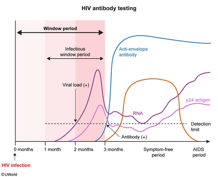
- HIV antibody assays (i.e., third-generation and below): Detect IgM and IgG antibodies.
- Laboratory methods
- Enzyme-linked immunosorbent assays (ELISA)
- HIV-1 and HIV-2 antibody differentiation immunoassay
- Laboratory-based test that can differentiate between HIV-1 and HIV-2 (provides separate results for each analyte)
- Most commonly used confirmatory test in the US
- Western blot: Detects only IgG antibody to HIV-1
- Laboratory methods
- Combination HIV antibody with HIV antigen test (i.e., fourth-generation and above): Can detect HIV IgG and IgM antibodies and p24 antigen.
- Cannot differentiate between HIV-1 and HIV-2 infection
Treatment
See HIV therapy
Prevention
- Screening:
- Universal for ages 15–65 yrs & all pregnant pts.
- Test: 4th-gen HIV-1/2 Ag/Ab assay (detects p24 Ag); confirm w/ Ab differentiation assay.
- PrEP (Pre-Exposure Prophylaxis):
- Candidates: HIV-negative patients w/ ongoing high-risk exposures (MSM, PWID, partner w/ unsuppressed VL).
- Regimens: Daily oral TDF/FTC (or TAF/FTC; not for receptive vaginal sex) OR q2mo Cabotegravir IM.
- Key Step: Must confirm HIV(-) status before starting (prevents drug resistance); check CrCl & HBV status.
- PEP (Post-Exposure Prophylaxis):
- Timing: Must be initiated A.S.A.P. (ideally < 2 hours, maximum 72 hours post-exposure); duration 28 days.
- Regimen: 3 drugs Dolutegravir (or Raltegravir) + TDF/FTC.
- PMTCT (Perinatal Transmission):
- Maternal ART: Immediate & universal for all HIV(+) pregnant pts.
- Delivery: Vaginal if VL 1000 copies/mL; Scheduled C-section at 38 wks + IV Zidovudine (AZT) if VL > 1000 copies/mL.
- Postpartum: Infant oral AZT prophylaxis; breastfeeding contraindicated in US/developed nations.
- TasP (Treatment as Prevention):
- U=U: Undetectable VL (< 50 copies/mL) for >6 mo = zero sexual transmission risk.
- Harm Reduction:
- Syringe exchange, Opioid Agonist Therapy (Methadone/Buprenorphine), prompt STI treatment.
Preventive health care
- Opportunistic Infection Prophylaxis:
- CD4 < 200: PCP -> TMP-SMX (Stop when CD4 > 200 for > 3 mo).
- CD4 < 100: Toxoplasma (if IgG (+)) -> TMP-SMX (Stop when CD4 > 200 for > 3 mo). c
- Toxoplasmic encephalitis (TE) in HIV-infected patients is almost exclusively caused by the reactivation of latent tissue cysts (bradyzoites) acquired in the past, rather than a new acute primary infection.
- CD4 < 150: Histoplasma (endemic areas) -> Itraconazole.
- CD4 < 50: MAC -> No prophylaxis if ART started immediately (Azithromycin only if ART delayed). c
- Prompt ART initiation rapidly restores immune function and lowers MAC risk without unnecessary drug exposure or resistance
- Immunizations:
- Contraindicated if CD4 < 200: Live vaccines (MMR, Varicella, LAIV nasal flu). c
- Influenza: Annual inactivated vaccine.
- Pneumococcal: PCV20 alone OR PCV15 + PPSV23 (≥8 wks later).
- Hepatitis B: High-dose 3-dose series if anti-HBs < 10 mIU/mL.
- Hepatitis A: 2-dose series if MSM, IVDU, or chronic liver disease.
- Meningococcal (MenACWY): 2-dose primary series + booster q5y.
- HPV: 3-dose series for ages 9–26 (can consider up to age 45).
- Herpes Zoster: Recombinant (Shingrix) 2 doses for all adults ≥18 yo.
- Infectious Disease Screening:
- Tuberculosis: TST or IGRA at diagnosis. ≥5 mm induration on TST = positive. If (+), perform CXR to r/o active TB before treating LTBI (Isoniazid x 9 mo). c
- Syphilis, Gonorrhea, Chlamydia: Baseline & annually (site-specific NAAT for GC/CT).
- Hepatitis B & C: Baseline serologies (annual HCV RNA screening if high-risk).
- Cancer & Routine Screening:
- Cervical Pap: At diagnosis -> annually x 3 -> q3y if normal.
- Anal Cancer: Annual digital anal rectal exam (DARE) ± anal Pap smear.
- Standard Cancers: Age-matched USPSTF screening for breast, colon, and lung.
- Metabolic & Bone Health:
- Lipids & Glucose: Baseline, 1–3 mo post-ART initiation, then annually.
- Statin Therapy: Moderate-intensity statin recommended for ages 40–75 regardless of baseline ASCVD risk.
- DEXA Scan: Screen men ≥50 yo and postmenopausal women.
HIV in pregnancy
- Antepartum Management:
- cART Initiation: Start immediately upon diagnosis regardless of CD4 count or VL. c
- Regimen: 2 NRTIs (e.g., Tenofovir + Emtricitabine or Abacavir + Lamivudine) + Integrase Inhibitor (e.g., Dolutegravir or Raltegravir).
- Avoid: Invasive prenatal diagnostic procedures (e.g., CVS, amniocentesis) unless maternal VL is undetectable.
- Intrapartum Management:
- VL ≤ 1,000 copies/mL at 36 weeks: Vaginal delivery indicated. Continue oral cART during labor.
- Transmission risk with cART is <1%, regardless of route of delivery
- VL > 1,000 copies/mL (or unknown) at 36 weeks: Scheduled Cesarean delivery at 38 weeks (prior to labor/ROM).
- Transmission risk during vaginal birth increases significantly
- Intrapartum IV Zidovudine (AZT): Administer continuously if VL > 1,000 copies/mL or unknown (start 3 hrs prior to C-section).
- Procedures to Avoid: Artificial rupture of membranes (AROM), fetal scalp electrodes/blood sampling, operative vaginal delivery (vacuum/forceps).
- VL ≤ 1,000 copies/mL at 36 weeks: Vaginal delivery indicated. Continue oral cART during labor.
- Postpartum & Infant Management:
- Infant Post-exposure Prophylaxis (PEP) (start within 6-12 hrs of birth):
- Low Risk (maternal VL <50 copies/mL): Zidovudine (AZT) for 4 weeks.
- High Risk (maternal VL >50 copies/mL or unknown): 3-drug cART regimen (AZT + Lamivudine + Nevirapine or Raltegravir) for 6 weeks.
- Infant Feeding: Breastfeeding is strictly contraindicated in high-resource settings (e.g., US) regardless of maternal VL; recommend formula feeding.
- Infant Post-exposure Prophylaxis (PEP) (start within 6-12 hrs of birth):