Epidemiology
- General: Approx. 90–95% of adults are EBV-seropositive worldwide.
- Peak incidence (of symptomatic disease): 15–24 years of age
Etiology
- Pathogen: Epstein-Barr virus (EBV), also called human herpesvirus 4 (HHV-4)
- Transmission: Infectious mononucleosis spreads via bodily secretions, especially saliva. Therefore, it is also called kissing disease.
Pathophysiology
Mnemonic
Infects B cells through CD21, “Must be 21 to drink Beer in a Barr”
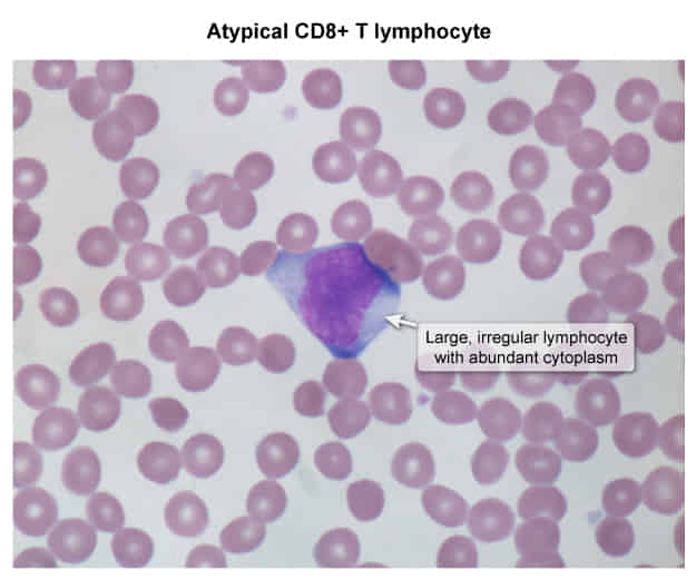
- EBV infects B lymphocytes in mucosal epithelium (e.g., oropharynx, cervix) via the CD21 receptor → infected B lymphocytes induce a humoral (B-cell) as well as a cellular (T-cell) immune response → an increased concentration of atypical lymphocytes in the bloodstream, which are CD8+ cytotoxic T cells that fight infected B lymphocytes
- Atypical lymphocytes are not infected by virus; they react to infected cells. They contain cytotoxic granules composed of perforin (creates holes in the infected cell’s membrane) and granzymes (enter the cytoplasm of infected cells and trigger cell death)
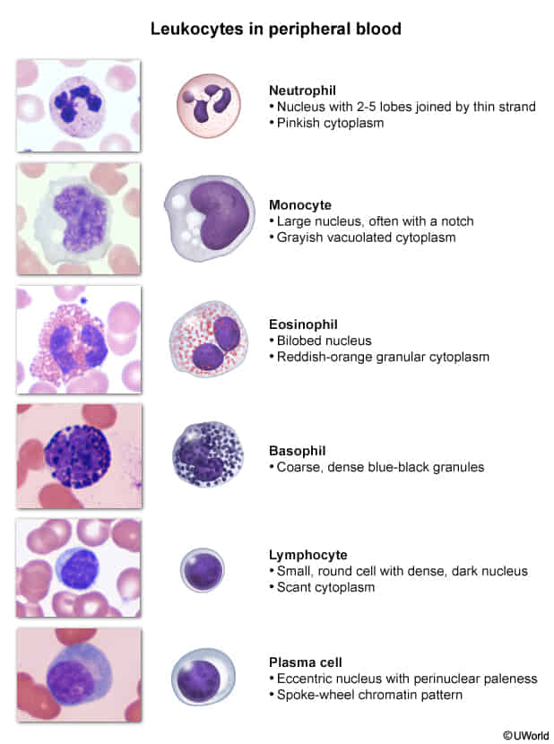
- The name “mononucleosis” is derived from the monocyte-like appearance of (atypical) lymphocytes under the microscope.
- The T-cell response is crucial for overcoming the infection. However, if the response is not sufficient (e.g., in HIV patients), B cells may proliferate uncontrollably and increase the risk of complications. For example, the high rate of proliferation increases the risk of oncogenic mutations that lead to the development of B-cell lymphomas.
Clinical features
- Classic Triad: Fever + Pharyngitis + Lymphadenopathy.
- Pharyngitis: Often severe, exudative, tonsillar enlargement.
- Lymphadenopathy: Posterior cervical chain (highly suggestive); can be generalized.
- Hepatosplenomegaly: Splenomegaly in >50% of cases.
- Fatigue: Profound, can last months.
- Rash: Maculopapular rash develops if patient is mistakenly treated with Amoxicillin or Ampicillin (mechanism is a hypersensitivity reaction, not a true penicillin allergy).
Warning
Splenomegaly can lead to a potentially life-threatening splenic rupture!
Diagnostics
- Monospot test
- Detects heterophile antibodies produced in response to EBV infection using RBCs from sheep or horses
- The mechanism remains unclear; EBV either induces a humoral response to heterophile antigens or stimulates nonspecific polyclonal activation of B cells.
- Patient’s serum is mixed with a solution of sheep/horse RBC in vitro
- Positive test: cross-reaction between heterophile antibodies and sheep/horse RBCs → agglutination
- Negative test
- No heterophile antibodies present → no cross-reaction → no agglutination
- In some cases, the test can show negative results if it is performed too soon (i.e. within the first 1–2 weeks after symptom onset) and antibodies have not developed yet. For this reason, a negative heterophile antibody test does not exclude the diagnosis of IM. Repeating the test after several days or checking anti-EBV IgM and IgG antibodies can help establish the diagnosis. c
- Specificity of ∼ 100%, sensitivity of 85%
- Detects heterophile antibodies produced in response to EBV infection using RBCs from sheep or horses
- Peripheral smear: lymphocytosis with > 10% atypical lymphocytes (in some cases, up to 90%)
- Since EBV typically causes a latent infection of B cells, only minimal amounts of EBV-specific antigens circulate in an immunocompetent host; therefore, blood tests for such antigens are insensitive to latent EBV infection.
Differential diagnostics
- Mononucleosis-like syndromes
- Streptococcal pharyngitis, tonsillitis
- Acute HIV infection
- Viral hepatitis
- CMV infection
- Toxoplasmosis
Treatment
- Primary / Supportive: Rest, fluids, NSAIDs/acetaminophen for fever/pain.
- Splenic Rupture Prevention: Strict avoidance of contact sports / strenuous exercise for >= 3-4 weeks (or until full symptom resolution). c
- Corticosteroids: NOT routinely indicated. Reserved for severe complications:
- Airway obstruction (massive tonsillar hypertrophy).
- Severe autoimmune hemolytic anemia or severe thrombocytopenia.
- Antivirals: Acyclovir/Valacyclovir NOT recommended (no clinical benefit). c
Complications
- Splenic Rupture: Rare, life-threatening emergency; presents with sudden severe LUQ pain, left shoulder pain (Kehr sign), hypotension/shock.
- Airway Obstruction: Upper airway compromise due to marked tonsillar enlargement.
- Hematologic: Cold agglutinin-mediated autoimmune hemolytic anemia (Coombs (+), IgM/C3d) and thrombocytopenia. c
- Neurologic: Guillain-Barré syndrome, encephalitis, transverse myelitis, facial nerve palsy.
- Malignancies (Long-term): Burkitt lymphoma (t(8;14)), Nasopharyngeal carcinoma, Hodgkin lymphoma, CNS lymphoma in immunocompromised (HIV).
Post-transplantation lymphoproliferative disorder
- Definition & Etiology:
- Proliferation of B cells driven by Epstein-Barr Virus (EBV) reactivation under chronic immunosuppression post-solid organ transplant (SOT) or HSCT.
- Highest Risk: Donor EBV (+) / Recipient EBV (-) status (D+/R-) and high-intensity immunosuppression (e.g., within 1st year post-transplant).
- Clinical Presentation:
- B-symptoms: Unexplained persistent fever, night sweats, weight loss.
- Lymphadenopathy (LAD): Painless, non-tender local/generalized LAD, tonsillar enlargement.
- Extranodal / Allograft Involvement:
- Allograft dysfunction (e.g., ↑ Cr in renal graft, ↑ LFTs in liver graft — mimics rejection).
- GI tract: Abdominal pain, GI bleeding, obstruction, or perforation.
- CNS: Focal deficits, AMS, headache.
- Diagnostic Workup:
- Initial/Screening: Quantitative EBV PCR (viral load) + ↑ Serum LDH + cytopenias on CBC.
- Imaging: Contrast CT (Neck/Chest/Abdomen/Pelvis) or FDG-PET/CT for staging and biopsy localization.
- Confirmatory / Gold Standard: Excisional lymph node or tissue biopsy demonstrating lymphoid proliferation, EBER positivity (EBV-encoded RNA via in situ hybridization), and CD20 markers.
- Key Differential:
- Acute Rejection: Infiltrates graft without systemic LAD/B-symptoms; biopsy shows T-cell or Ab-mediated rejection (EBER negative).
- CMV / Opportunistic Infection: Differentiated by specific PCRs, cultures, and absence of malignant clonal proliferation on biopsy.
- Management Hierarchy:
- Step 1 (First-line): Reduction of Immunosuppression (RI) — restores host T-cell surveillance (must monitor for acute allograft rejection).
- Step 2 (CD20+ / Persistent disease): Rituximab (anti-CD20 monoclonal antibody).
- Step 3 (Refractory / Aggressive monomorphic lymphoma): Systemic Chemotherapy (R-CHOP regimen).
- Adjunct: Surgery/radiation only for acute emergencies (e.g., bowel perforation, impending airway obstruction).