Epidemiology
Etiology
- Viral: Most common cause (~70-90%). Adenovirus, Rhinovirus, Coronavirus, EBV (Mononucleosis), CMV, HSV.
- Bacterial: Group A -hemolytic Streptococcus (GAS/S. pyogenes) is the most important bacterial cause (15-30% in children). t
- Other Bacterial: Fusobacterium necrophorum (Lemierre syndrome), N. gonorrhoeae, C. diphtheriae (rare/unvaccinated).
Pathophysiology
Clinical features
Acute bacterial tonsillopharyngitis
- Sudden onset of symptoms: fever, sore throat, dysphagia
- Significantly inflamed pharynx
- Pharyngeal and/or tonsillar erythema and edema
- Pharyngeal and/or tonsillar exudates (rare in children < 3 years of age)
- Fibrin deposits appear as white spots on the tonsils (杨梅舌). t
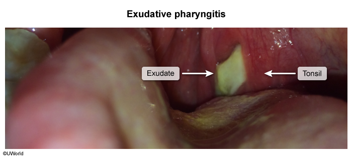
- See Gray-white exudates of throat
- Fibrin deposits appear as white spots on the tonsils (杨梅舌). t
- Palatal petechiae
- Cervical lymphadenitis
- Absence of cough
Acute viral tonsillopharyngitis
Accompanied by symptoms of a common cold (rhinitis and/or pharyngitis)
Diagnostics
- Centor / Modified McIsaac Criteria (Age 3–14: +1; 15–44: 0; ≥45: -1):
- Absence of cough (+1).
- Tender anterior cervical LAD (+1).
- History of fever / T > 38°C (+1).
- Tonsillar exudates or swelling (+1).
- Initial / Screening:
- Centor score 0–1: No diagnostic testing or Abx indicated (symptomatic care).
- Centor score ≥2: Perform Rapid Antigen Detection Test (RADT). c
- RADT: Highly specific (~95%), moderate sensitivity (~70–90%).
- Confirmatory / Gold Standard: Throat Culture.
- Mandatory in children/adolescents if RADT is negative (to prevent ARF). c
- Not routinely required in adults if RADT is negative due to low risk of ARF.
- Key Labs:
- Heterophile antibody test (Monospot) / EBV serologies if infectious mononucleosis suspected.
- Antistreptolysin O (ASO) titer
- Elevated levels suggest a previous GAS infection; not helpful to diagnose acute pharyngitis
- Indicated in the workup of nonsuppurative complications of GAS tonsillopharyngitis (e.g., acute rheumatic fever, PSGN).
Treatment
Complications
Infectious thrombophlebitis of the internal jugular vein (Lemierre syndrome)
- Etiology: Fusobacterium necrophorum (anaerobic GNR) following pharyngitis, dental infection, or tonsillitis in young, healthy pts (age 15–24). c
- Clinical Presentation:
- Oropharyngeal infection: Preceding sore throat/tonsillitis -> high fevers, rigors.
- Neck findings: SCM tenderness and induration along the internal jugular vein (IJV). c
- Septic pulmonary emboli: Dyspnea, pleuritic CP, hemoptysis, nodular lung lesions.
- Diagnosis:
- CT neck w/ IV contrast: Best initial test (shows IJV thrombosis, parapharyngeal swelling).
- CXR / CT chest: Bilateral nodular opacities, cavitary lesions, pleural effusions.
- Blood cultures: Confirmatory for F. necrophorum.
- Management:
- IV Abx: Empiric anaerobic coverage w/ Ampicillin-sulbactam OR Ceftriaxone + Metronidazole (3–6 wks).
- Anticoagulation: NOT routine (reserved for thrombus progression/persistent emboli despite 48–72h of Abx).
- Drainage: For persistent deep neck abscesses or empyema.
- Key Distractors:
- Peritonsillar abscess: Uvula deviation, trismus, “hot potato” voice; no IJV thrombosis or lung emboli.
- Tricuspid endocarditis: IVDU history, holosystolic murmur; no preceding neck pain/IJV clot.