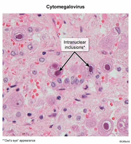
Often asymptomatic or presents as a mononucleosis-like syndrome.
Symptoms include fever, malaise, myalgias, and atypical lymphocytosis.
A key distinguishing feature from EBV mononucleosis is the negative heterophile antibody (Monospot) test. Pharyngitis and significant lymphadenopathy are less common than in EBV.
At high risk for severe, end-organ disease, particularly when the CD4+ count is <50 cells/µL in AIDS patients.
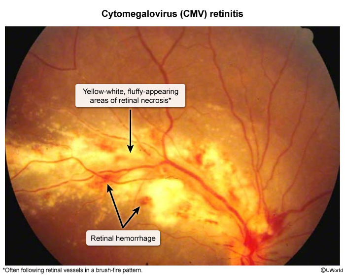
Retinitis: The most common manifestation in advanced AIDS, presenting with hemorrhage and cotton-wool exudates on fundoscopy (“pizza pie” or “ketchup and mustard” appearance). Can lead to retinal detachment and blindness.
Colitis: Results in diarrhea, fever, and abdominal pain. Endoscopy often reveals ulcerations. c
Esophagitis: Causes odynophagia (painful swallowing). Endoscopy typically shows large, linear ulcers in the distal esophagus.
Pneumonitis: A significant cause of morbidity and mortality in transplant recipients, especially after lung or hematopoietic stem cell transplants.
Encephalitis: Can occur in severely immunocompromised patients.
Congenital CMV
The most common congenital viral infection.
~90% of infected newborns are asymptomatic at birth.
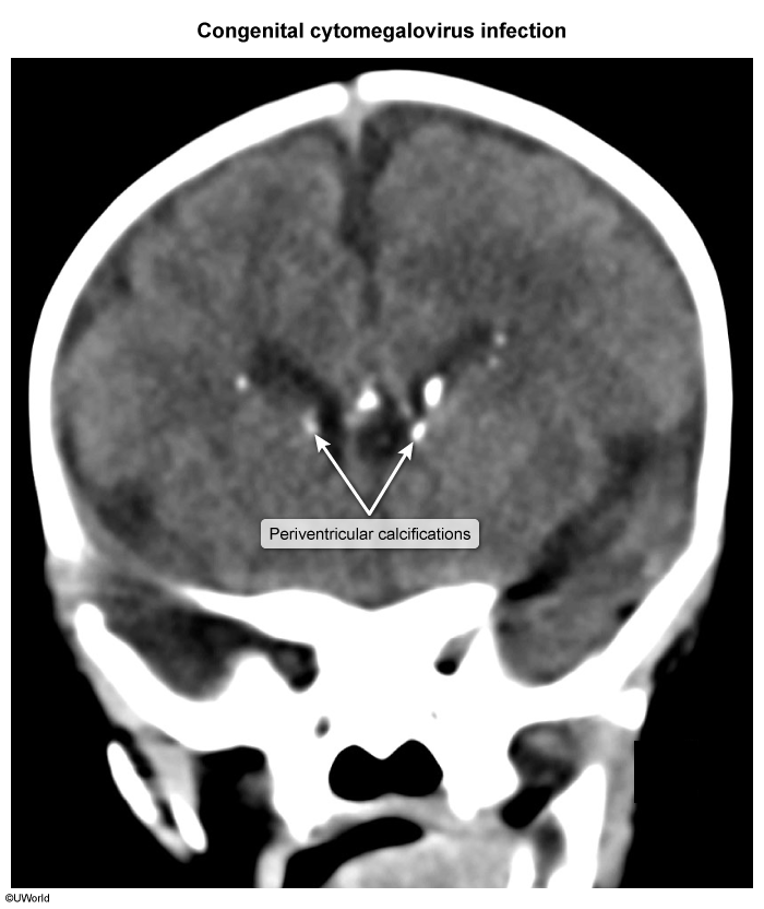
Symptomatic newborns may present with hepatosplenomegaly, jaundice, thrombocytopenic purpura (“blueberry muffin” rash), microcephaly, and periventricular calcifications.
CMV is a leading cause of non-hereditary sensorineural hearing loss in children.
Diagnostics
Initial / Screening Tests:
Immunocompetent: Monospot test = Negative; peripheral smear shows atypical lymphocytes.
Immunocompromised: CMV Quantitative PCR (plasma/blood) or viral load monitoring.
Congenital: CMV PCR in urine or saliva within first 2–3 weeks of life.