- Description
- Pancytopenia caused by bone marrow insufficiency
- Should not be confused with aplastic crisis, a condition in which erythropoiesis is temporarily suppressed (e.g., due to parvovirus B19 infection in patients with hemolytic anemias)
- Etiology
- Idiopathic in > 50% of cases
- Possibly immune-mediated
- May follow acute hepatitis (hepatitis-associated aplastic anemia)
- Medication side effects: carbamazepine, methimazole, NSAIDs, chloramphenicol, propylthiouracil, sulfa drugs, cytostatic drugs (esp. alkylating agents and antimetabolites)
- Dose-dependent reactions are caused by medications (eg, chemotherapy, immunosuppressives) that are toxic to the bone marrow when drug levels exceed a certain concentration for a certain period. They tend to be predictable in onset.
- Idiosyncratic reactions are often caused by antiseizure medications (eg, carbamazepine, valproic acid), sulfonamides, or nifedipine. They are unpredictable, unrelated to the dose of the drug, and may occur at any point during or after therapy. Although the mechanism is unclear, patients often have genetic mutations in drug-metabolizing enzymes or efflux pumps, which may promote toxicity.
- Toxins: benzene, cleaning solvents, insecticides, toluene
- Ionizing radiation
- Viruses: HBV, EBV, CMV, HIV
- Fanconi anemia
- Idiopathic in > 50% of cases
- Pathophysiology
- Multipotent hematopoietic stem cells are destroyed by cytotoxic T cells or direct cytotoxic injury → bone marrow aplasia/hypoplasia → lack of circulating peripheral blood cells
- Diagnostics
- Laboratory studies, e.g.:
- CBC: pancytopenia (in contrast to aplastic crisis characterized by anemia only), or normocytic or macrocytic anemia
- Reticulocyte count: low
- Erythropoietin level: high
- Bone marrow findings: hypocellular with prominent fat spaces (“empty marrow” appearance)
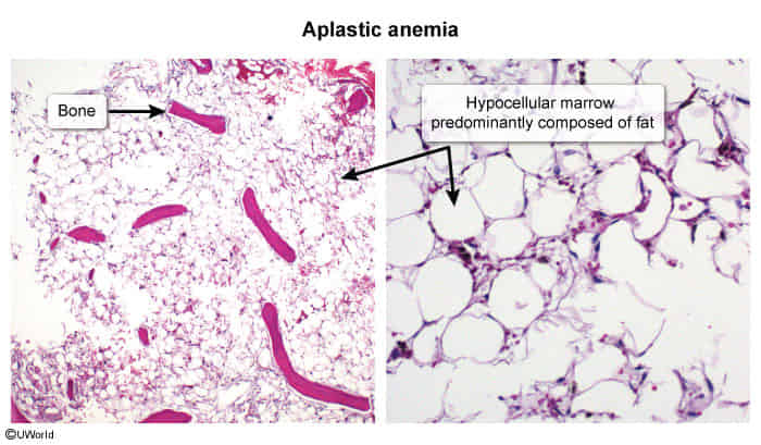
- Laboratory studies, e.g.:
Differential diagnostics
Feature Myelofibrosis (MF) Aplastic Anemia (AA) Pathophysiology Myeloproliferative Neoplasm (MPN) → ↑ Cytokines → Reactive BM fibrosis Autoimmune destruction of hematopoietic stem cells (HSCs) → BM failure BM Biopsy Hypercellular → Fibrotic. “Dry tap” common. Markedly hypocellular / empty; replaced by fat. Splenomegaly Massive (due to Extramedullary Hematopoiesis - EMH). Hallmark finding. Absent. Peripheral Smear Leukoerythroblastosis & Dacrocytes (teardrop RBCs). Pancytopenia with normal morphology. Genetics JAK2, CALR, MPL mutations (~90%). No specific mutations; idiopathic/autoimmune. Key Sx Constitutional “B” symptoms, early satiety, bone pain. Sx of pancytopenia: fatigue, infections, bleeding. Age Typically >50 y/o. Bimodal: young adults & elderly.
Link to originalMyelofibrosis vs Aplastic anemia
- Myelofibrosis: splenomegaly
- Evidence of extra medullary hematopoesis
- Aplastic anemia: no
- Explanation: In myeloproliferative disorders like myelofibrosis, niche in marrow is disturbed by slow fibrosis of marrow so stem cells migrate and extra medullary haematopoiesis occurs. But in aplastic anemia (whether primary or secondary to drugs etc) stem cells themselves are destroyed or affected so haematopoiesis not possible anywhere. So we don’t get hepatosplenomegaly and other evidences of extramedullary haematopoiesis.
- Myelofibrosis: splenomegaly
- Treatment
- Consider need for supportive care, e.g.:
- Platelet transfusions, and/or blood transfusions
- Bone marrow stimulants (e.g., GM-CSF, eltrombopag)
- Manage the underlying cause (e.g., cessation of the causative agent, or treatment of infections)
- If no cause is identified, treatment for idiopathic aplastic anemia may include:
- Immunosuppressive therapy to prevent further autoimmune marrow destruction
- Cyclosporine
- Antithymocyte globulin
- Consider hematopoietic stem cell transplantation in patients under 50 years of age with a suitable stem cell donor.
- Immunosuppressive therapy to prevent further autoimmune marrow destruction
- Consider need for supportive care, e.g.:
Fanconi anemia
- Etiology & Pathophysiology:
- AR/X-linked defect in DNA cross-link repair (FANC mutations) leading to chromosomal instability.
- Most common inherited cause of aplastic anemia; typical presentation at 5–10 years of age.
- Clinical Presentation:
- Pancytopenia: Bleeding/petechiae (thrombocytopenia is often 1st sign), fatigue/pallor (anemia), recurrent infections (leukopenia).
- Congenital Anomalies:
- Skeletal: Hypoplastic or absent thumbs, radial ray defects, short stature. c
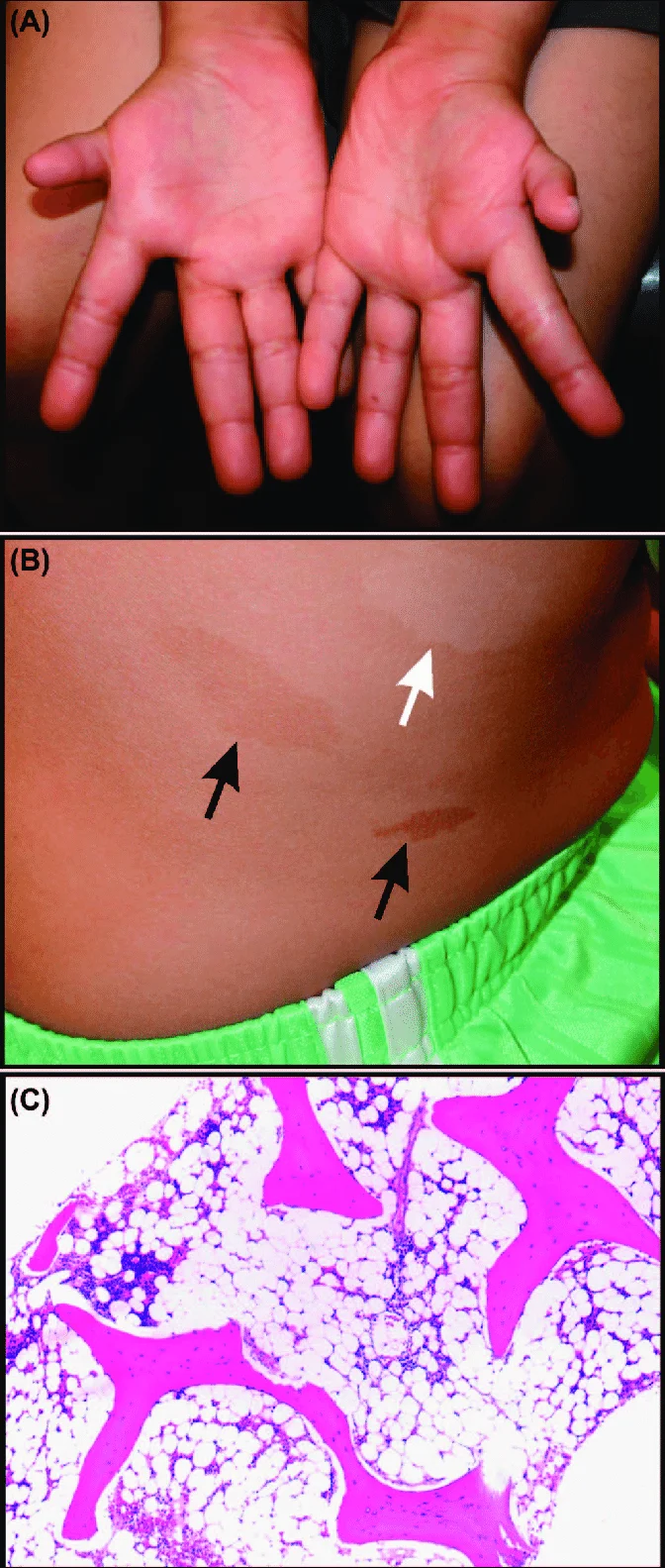
- Skin: Café-au-lait spots, hyper-/hypopigmented macules.
- Other: Microcephaly, horseshoe kidney, developmental delay, hypogonadism.
- Diagnostic Workup:
- Initial Labs: Pancytopenia with macrocytosis (↑ MCV) and ↑ HbF. c
- Confirmatory (Gold Standard): Chromosomal breakage analysis exposed to diepoxybutane (DEB) or mitomycin C (MMC).
- BM Biopsy: Marked hypocellularity with fatty replacement.
- High-Yield Differentials:
- Diamond-Blackfan Anemia: Pure RBC aplasia (normal WBC/PLT), triphalangeal thumbs, presents in infancy (<1 yo).
- Acquired Aplastic Anemia: Normal thumbs/no congenital anomalies, negative chromosomal breakage.
- Management:
- Definitive/Curative: Allogeneic HSCT (reduced-intensity conditioning).
- Medical / Bridge: Synthetic androgens (oxymetholone), G-CSF, irradiated/leukodepleted blood products.
- Key Step 2 Caution: Avoid ionizing radiation and alkylating chemotherapy.
- Major Complications:
- Hematologic: AML and MDS.
- Solid tumors: Squamous cell carcinoma (SCC) of head, neck, and GI/GU tract.