Family: Herpesviridae (enveloped, double-stranded, linear DNA virus).
Latency: A key feature. After primary infection, the virus becomes latent in sensory nerve ganglia and can reactivate later.
HSV-1: Latent in the trigeminal ganglia.
HSV-2: Latent in the sacral ganglia.
Histology: Look for Cowdry type A intranuclear inclusions and multinucleated giant cells on a Tzanck smear.
HSV-infected cells express viral glycoproteins (e.g., gB, gH/gL) on their surface.
These glycoproteins are fusogenic, meaning they induce the cell membrane of the infected cell to fuse with the membranes of adjacent, uninfected cells.
This process of cell fusion creates a single, large cell membrane containing multiple nuclei, forming a multinucleated giant cell (syncytium).
Clinical Presentations
Although HSV-1 is classically “above the waist” and HSV-2 is “below the waist,” either type can infect either location.
HSV-1 Manifestations:
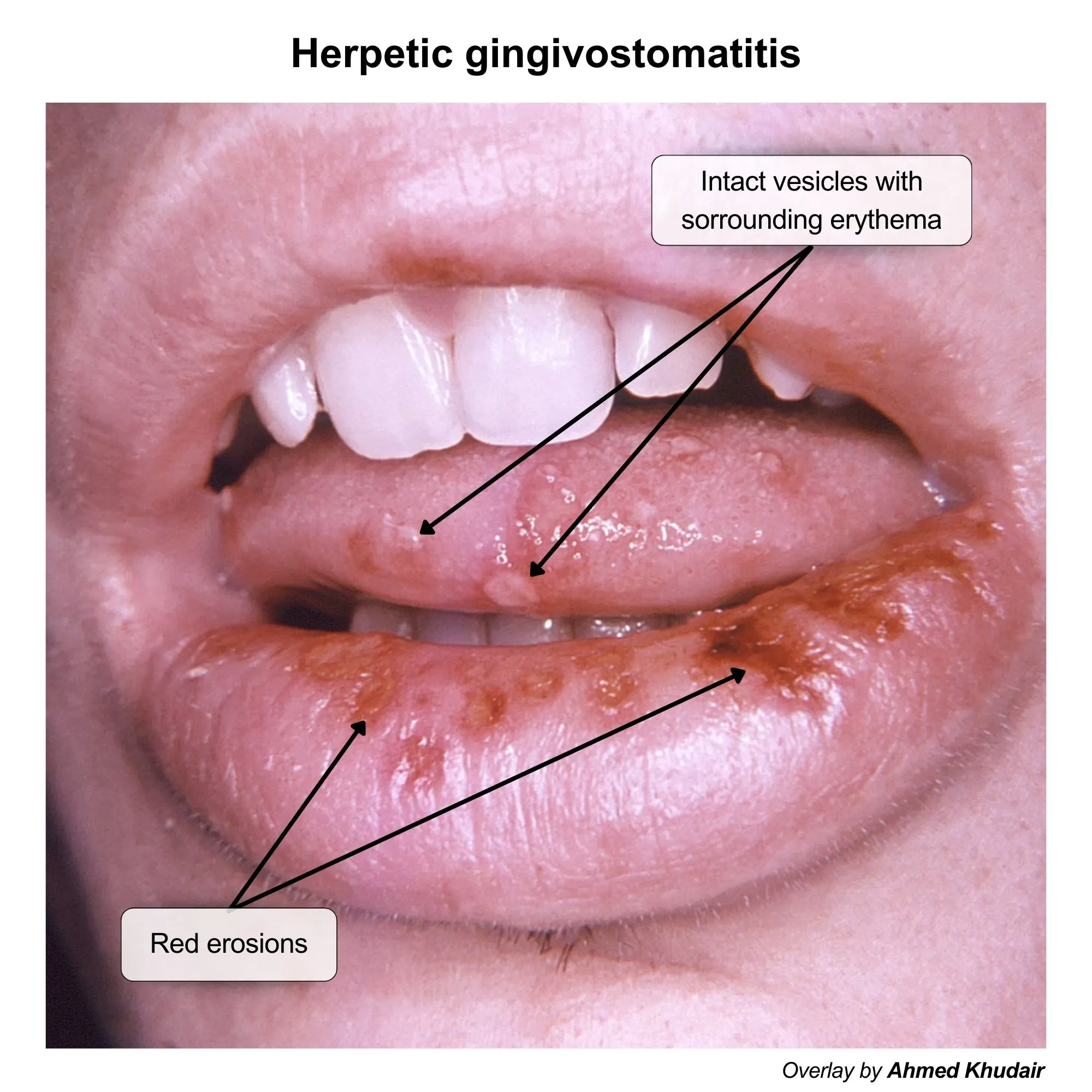
Gingivostomatitis: Most common primary HSV-1 manifestation in children; painful vesicles on oral mucosa, gingiva, tongue, and perioral skin + high fevers, drooling, refusal to eat/drink.
Herpes Labialis (Cold Sores): Recurrent vesicular lesions on or around the lips.
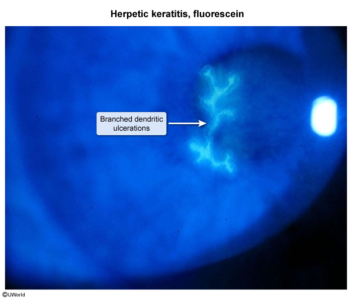
Keratoconjunctivitis: Corneal infection that can cause dendritic ulcers visible with fluorescein stain; a major cause of blindness. c
Unilateral in immunocompetent; bilateral in immunocompromised
Temporal Lobe Encephalitis: The most common cause of sporadic, fatal encephalitis. Presents with fever, headache, altered mental status, seizures, and personality changes.
Findings:Multinucleated giant cells, Cowdry A inclusions.
Differential (HIV):
CMV: Large, linear ulcers; inclusions at ulcer base.
Candida: White plaques/pseudomembranes.
HSV-2 Manifestations:
Genital Herpes: Causes painful vesicular lesions on the genitals and perineum, often with inguinal lymphadenopathy. Recurrences are more frequent and severe with HSV-2 than with genital HSV-1.
Aseptic Meningitis: A more common complication with HSV-2 than with HSV-1.
Other Manifestations (Either HSV-1 or HSV-2):
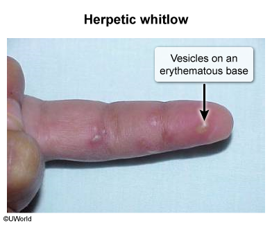
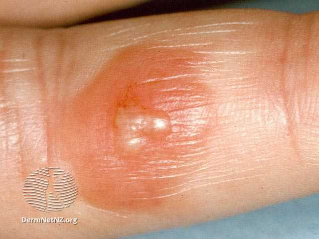
Herpetic Whitlow: A painful vesicular infection of the finger, common in healthcare workers or children who suck their thumbs. c
Erythema Multiforme: An acute, immune-mediated condition characterized by “target lesions,” often triggered by a preceding HSV infection.
Neonatal Herpes: A severe, often life-threatening infection acquired during delivery. It can be localized to the Skin, Eyes, and Mouth (SEM), involve the CNS, or be a disseminated multi-organ disease.
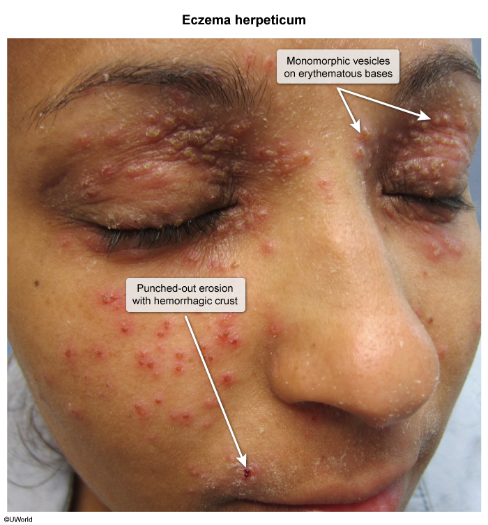
Eczema herpeticum: a potentially life-threatening dermatologic emergency caused by secondary superinfection with HSV (usually HSV-1) superimposed on pre-existing skin barrier dysfunction, most commonly atopic dermatitis (AD). Associated with systemic signs: fever, irritability, and regional lymphadenopathy (LAD).
Diagnosis
PCR: The gold standard for diagnosis, especially for CNS infections (analyzing CSF) or direct testing of lesions.
Viral Culture: Can be used to isolate the virus from an active lesion.
Tzanck Smear: A classic (but less sensitive/specific) test showing multinucleated giant cells and intranuclear inclusions.
Serology: Detects antibodies and can differentiate between HSV-1 and HSV-2; useful for confirming chronic infection in patients without active lesions.
Management
Mechanism: Antivirals like Acyclovir, Valacyclovir, and Famciclovir are guanosine analogs that inhibit viral DNA polymerase.
Indications:
Primary Infection: Treatment can shorten the duration and severity of the first outbreak.
Episodic Therapy: Used to manage recurrent outbreaks.
Suppressive Therapy: Daily low-dose antiviral medication is used for patients with frequent (e.g., >6 per year) or severe recurrences to reduce their frequency.
Severe Infections: HSV encephalitis or neonatal herpes requires immediate IV Acyclovir. Untreated HSV encephalitis has a high mortality rate.
Complications
Secondary Bacterial Superinfection: S. aureus cellulitis or bacteremia (especially in eczema herpeticum). c
HSV in Pregnancy
Transmission Risk: Highest with primary 3rd trimester infection (30–50%); lowest with recurrent infection (<1%).
Diagnosis: HSV PCR from active lesion swab (gold standard).
Prophylaxis: Acyclovir or Valacyclovir PO starting at 36 weeks until delivery for any pt with a history of genital HSV. c
Delivery Decision:
Active lesions OR prodromal symptoms at labor →\rightarrow→ Cesarean section.
No active lesions AND no prodrome at labor →\rightarrow→ Vaginal delivery (safe even w/ history of HSV).