Epidemiology
- Women: 15–44 years
Etiology
- Genetic predisposition
- HLA-DR2 and HLA-DR3 are commonly present in individuals with SLE.
- Genetic deficiency of classical pathway complement proteins (C1q, C2, C4) in approx. 10% of affected individuals
- Hormonal factors: Hyperestrogenic states (e.g., due to oral contraceptive use, postmenopausal hormonal therapy, endometriosis) are associated with an increased risk of SLE.
- Environmental factors
- Cigarette smoking and silica exposure increase the risk of developing SLE.
- UV light and EBV infection may trigger disease flares
- Ultraviolet rays and sun exposure lead to increased cell apoptosis
- Drugs such as procainamide or hydralazine (see “Drug-induced lupus erythematosus”)
Pathophysiology
Hypotheses:
- Autoantibody development: deficiency of classical complement proteins (C1q, C4, C2) → failure of macrophages to phagocytose immune complexes and apoptotic cell material (i.e., plasma and nuclear antigens) → dysregulated, intolerant lymphocytes targeting normally hidden intracellular antigens → autoantibody production (e.g., ANA, anti-dsDNA)
- Normally, apoptotic cells are engulfed by macrophages during apoptosis, avoiding the release of intracellular content that induces inflammation or an immune response in the extracellular environment.
- Autoimmune reactions
- Type III hypersensitivity (most common in SLE) → antibody-antigen complex formation in microvasculature → complement activation and inflammation → damage to skin, kidneys, joints, small vessels
- Type II hypersensitivity → IgG and IgM antibodies directed against antigens on cells (e.g., red blood cells) → cytopenia
Clinical features
- Classic triad: fever, joint pain, and rash in a woman of childbearing age.
- Highly variable presentation, known as “the great imitator”.
Common
- Constitutional: fatigue, fever, weight loss
- Joints (> 90% of cases)
- Arthritis and arthralgia c
- Distal symmetrical polyarthritis: most commonly affects the joints of the fingers, carpal joints, and the knee
- Skin (85% of cases)
- Malar rash: Fixed, erythematous maculopapular rash over the bridge of the nose and cheeks, sparing the nasolabial folds.
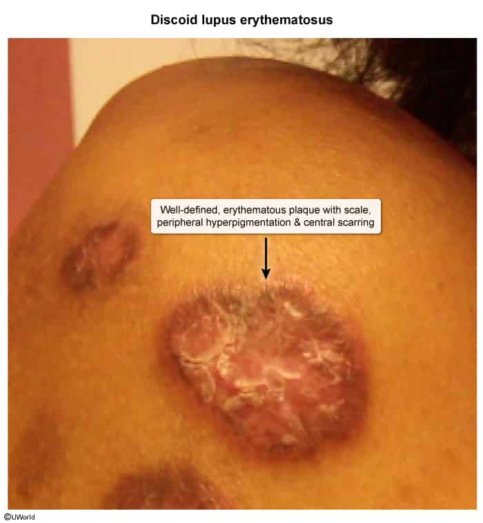
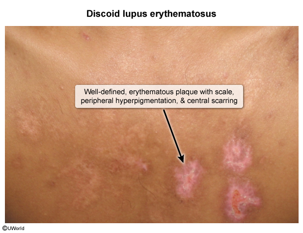
- Discoid rash: Scarring, erythematous plaques with central atrophy and follicular plugging. c
- Photosensitivity: Erythematous eruption following sunlight exposure.
- Ultraviolet rays and sun exposure lead to increased cell apoptosis
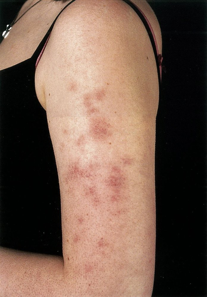
- Ultraviolet rays and sun exposure lead to increased cell apoptosis
- Painless oral or nasopharyngeal ulcers.
- Non-scarring alopecia.
- Raynaud phenomenon c
- Serositis: Pleuritis (pleuritic chest pain), pericarditis. t
Tip
Both rheumatoid arthritis and SLE arthritis affect the MCP and PIP joints, but SLE does not usually lead to deformities.
Less common
- Hematological: petechiae, pallor, or recurrent infections due to cytopenias
- Warm AIHA, IgG causes extravascular hemolysis.
- Kidneys: nephritis with proteinuria (see “Lupus nephritis”)
- Mesangial and/or subendothelial deposition of immune complexes (e.g., anti-dsDNA antibodies, anti-Sm antibodies) → expansion and thickening of mesangium, capillary walls, and/or glomerular basement membrane
- Heart
- Serositis (pleuritis, pericarditis w/ friction rub or effusion).
- Libman-Sacks endocarditis (sterile verrucous vegetations on mitral/aortic valves). c
- Increased risk of premature CAD/MI.
- Neuropsychiatric:
- Seizures, lupus psychosis (rule out steroid-induced psychosis), transverse myelitis, peripheral neuropathy, and cognitive impairment. c
Special clinical manifestations
Discoid lupus erythematosus
- Epidemiology & Triggers
- Demographics: F > M, 20–40 years of age, highest prevalence in African Americans.
- Triggers: UV light, cigarette smoking (reduces antimalarial efficacy).
- Prognosis: Primarily cutaneous; 5–10% progress to systemic lupus erythematosus (SLE).
- Clinical Features
- Distribution: Sun-exposed head and neck (scalp, face, conchal bowl of ear).
- Triad: Erythema, hyperkeratosis, and atrophy.
- Morphology: Indurated erythematous plaques with adherent scale; scale removal reveals keratotic spikes (“carpet tack sign”).
- Late stage: Central depigmented/atrophic scarring with hyperpigmented, active borders.
- Scalp: Permanent, irreversible scarring (cicatricial) alopecia.
Diagnostics
Laboratory studies
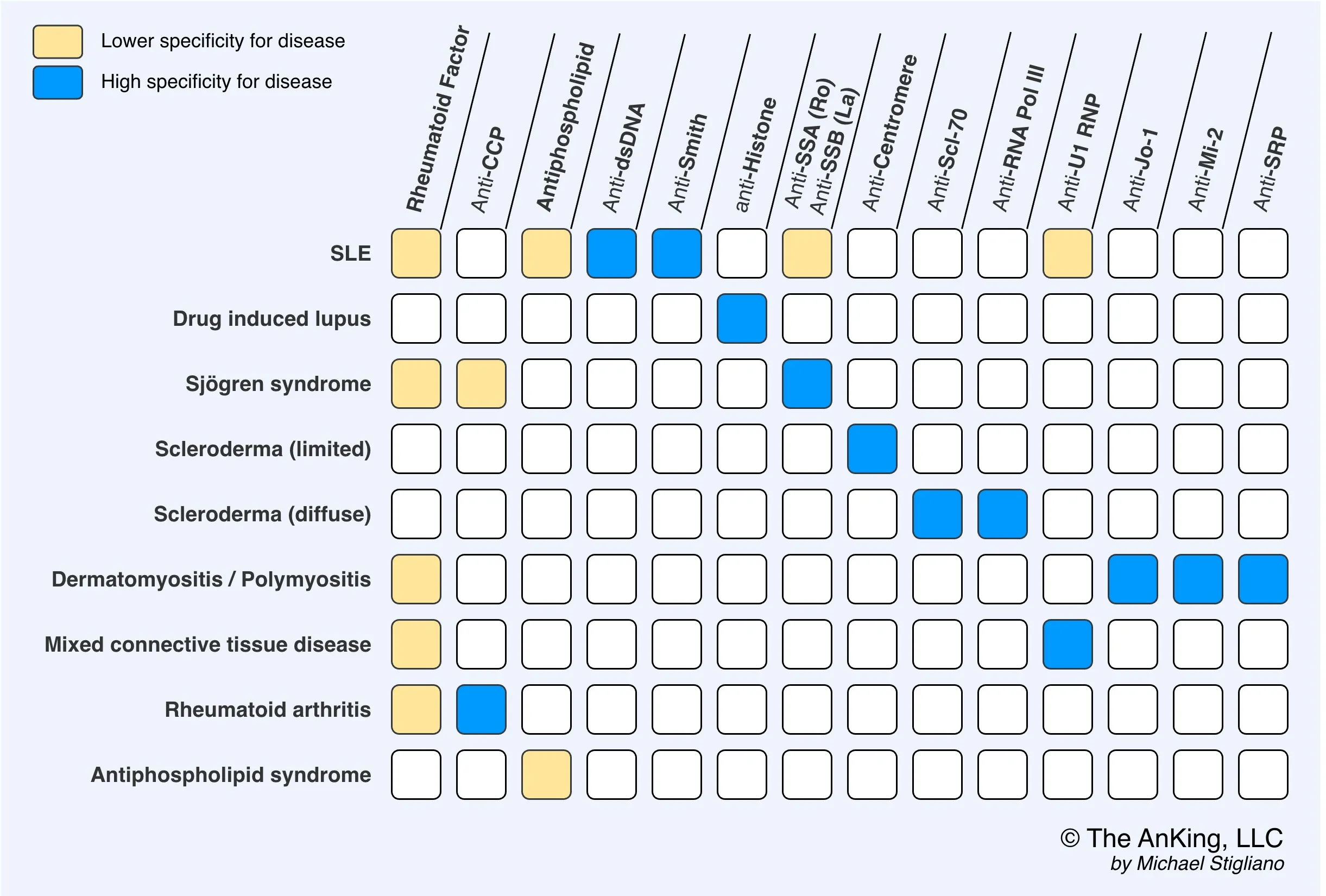
- Best initial test: ANA (Antinuclear Antibody) - highly sensitive (>95%) but not specific.
- Most specific antibodies: Anti-dsDNA and Anti-Sm (Anti-Smith).
- Anti-dsDNA antibodies
- Autoantibodies against double-stranded DNA
- Positive in 60–70% of patients
- Highly specific for SLE
- Levels correlate with disease activity (especially lupus nephritis activity).
- Anti-Sm antibodies
- Autoantibodies against Smith antigens (nonhistone nuclear proteins)
- Smith can bond to snRNAs to form snRNPs, which can form a spliceosome.
- Positive in < 30% of patients, but highly specific for SLE
- Autoantibodies against Smith antigens (nonhistone nuclear proteins)
- Anti-dsDNA antibodies
- Antiphospholipid antibodies: Screen all patients for antiphospholipid syndrome.
- Laboratory markers of disease activity and/or organ damage in SLE
- Complement levels: ↓ C3 and/or ↓ C4 in patients with active disease, factor B levels remain normal
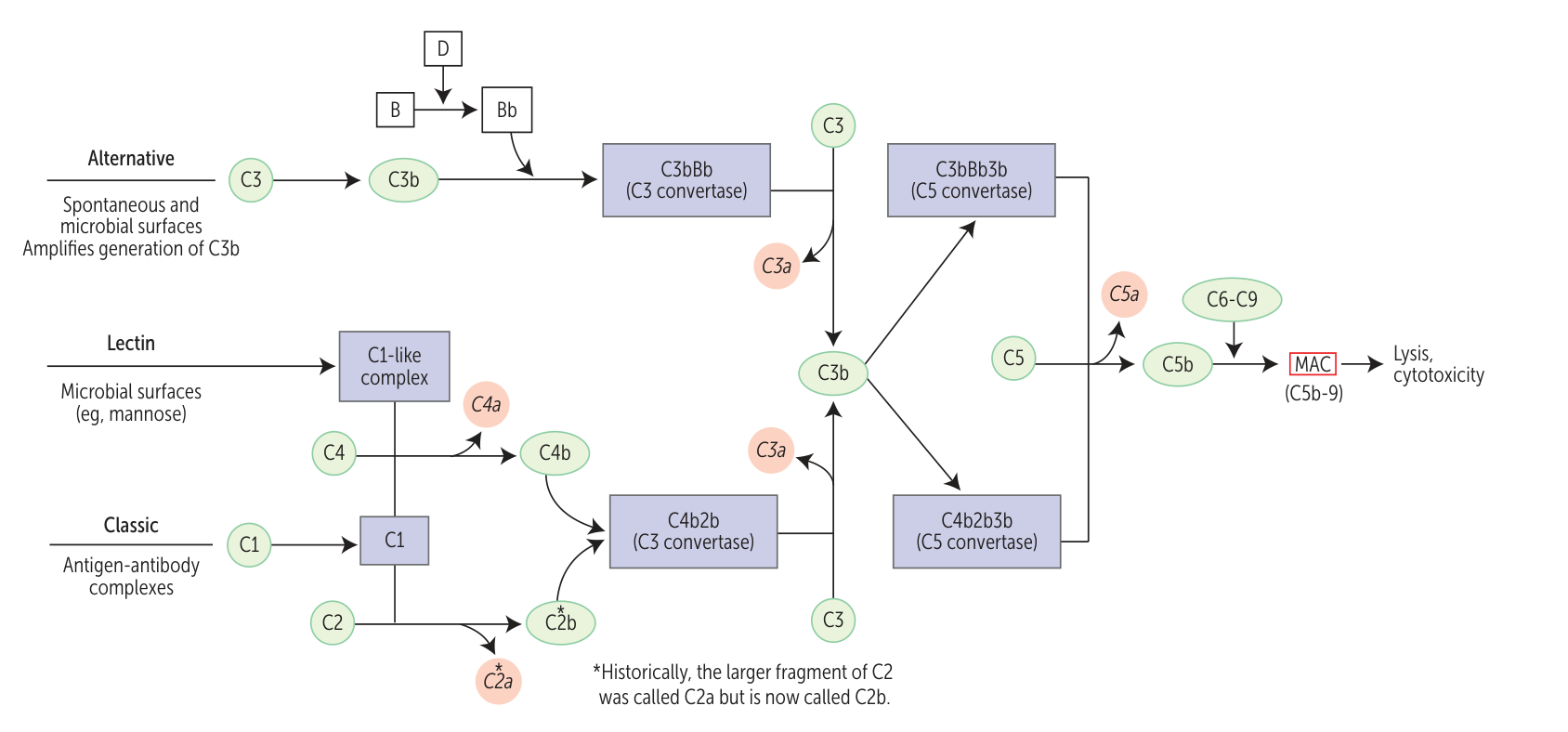
- Antigen-antibody complexes trigger classic pathway, which decreases C3 and C4. But factor B in alternative pathway is intact.
- Inflammatory markers
- ESR: may be elevated in patients with active disease
- CRP: often normal (may be elevated in patients with serositis, arthritis, or infections)
- CBC: may show leukopenia, thrombocytopenia, and/or autoimmune hemolytic anemia or anemia of chronic disease c2
- CMP: may show ↑ BUN and/or creatinine, and/or electrolyte abnormalities
- Urinalysis and urine microscopy: may show proteinuria, hematuria, and/or urinary casts
- Complement levels: ↓ C3 and/or ↓ C4 in patients with active disease, factor B levels remain normal
Tip
RPR and VDRL are usually used to test for syphilis but may also be positive in SLE. This happens in antiphospholipid syndrome as well.
Skin biopsy
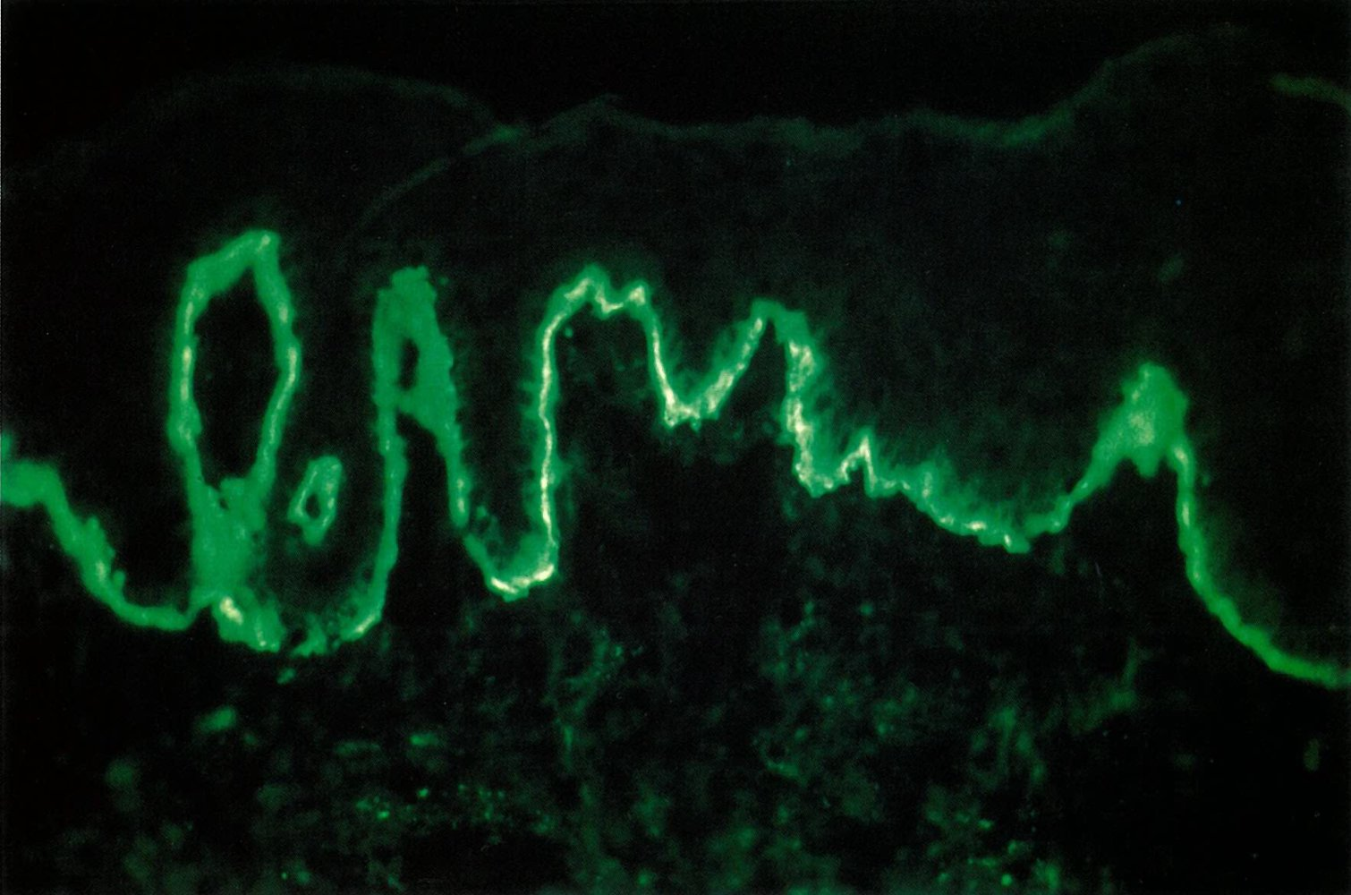
- Lupus band test (LBT): a direct immunofluorescence staining technique used to detect immunoglobulin and complement component deposits along the dermoepidermal junction in affected and unaffected skin in patients with SLE
EULAR/ACR 2019 Criteria (≥10 Points Required)
- Must include ≥1 clinical criterion. Count only the highest score per domain.
- Clinical Domains:
- Renal: Biopsy Class III/IV LN (10 pts), Class II/V LN (8 pts), Proteinuria >0.5g/24h (4 pts).
- Skin: Malar/acute rash (6 pts), Discoid/subacute (4 pts), Alopecia or oral ulcers (2 pts).
- Musculoskeletal: Arthritis (6 pts).
- Serosal: Pericarditis (6 pts), Pleural/pericardial effusion (5 pts).
- Neuro: Seizures (5 pts), Psychosis (3 pts), Delirium (2 pts).
- Heme: Hemolytic anemia (4 pts), Thrombocytopenia (4 pts), Leukopenia (3 pts).
- Fever: >38°C (2 pts).
- Immunologic Domains:
- Antibodies: Anti-dsDNA or Anti-Sm (6 pts), Antiphospholipid Ab (2 pts).
- Complement: Low C3 AND C4 (4 pts), Low C3 OR C4 (3 pts).
Treatment
- General: Sun protection, smoking cessation.
- Mild disease (skin, joints): Hydroxychloroquine (for all patients), NSAIDs, low-dose corticosteroids.
- Hydroxychloroquine: decreases complement-dependent antigen-antibody reactions.
- Moderate disease (serositis, significant cytopenias): Add moderate-dose corticosteroids, possibly immunosuppressants like azathioprine or methotrexate.
- Severe disease/Lupus Nephritis: High-dose corticosteroids plus cyclophosphamide or mycophenolate mofetil.
Complications
- End-Stage Renal Disease (ESRD): Resulting from untreated or refractory proliferative LN.
- Accelerated Atherosclerosis: Cardiovascular disease is the leading cause of death in long-standing SLE.
- Antiphospholipid Syndrome (APS): Hypercoagulability leading to arterial/venous thrombosis and recurrent pregnancy loss. c
- Neonatal Lupus: Occurs in infants of mothers with anti-Ro/SSA or anti-La/SSB; can cause permanent congenital third-degree heart block and transient cutaneous lesions.
- Avascular Necrosis (AVN): Often of the femoral head, secondary to prolonged high-dose corticosteroid use.
- Infections: Due to immunosuppressive therapy and intrinsic immune dysfunction (leading cause of death in early disease).
Cardiovascular disease
- ↑ Risk of thrombosis in all patients with SLE (especially if secondary antiphospholipid syndrome is present)
- ↑ Risk of myocardial infarction and stroke because of accelerated Atherosclerotic cardiovascular disease
Special patient groups
Neonatal lupus syndrome
- Etiology & Pathophysiology:
- Transplacental transfer of maternal anti-SSA/Ro and anti-SSB/La IgG autoantibodies.
- Mother may have diagnosed SLE, Sjögren syndrome, or be completely asymptomatic.
- Clinical Features:
- Cardiac (permanent, irreversible):
- Congenital 3rd-degree (complete) AV block: Presents as persistent fetal/neonatal bradycardia (HR < 100 bpm). c
- Cardiomyopathy, CHF, or endocardial fibroelastosis.
- Cutaneous (transient, self-resolves by ~6–8 months as maternal IgG wanes):
- Annular, erythematous scaly plaques with central clearing.
- Classically involves face/scalp with periorbital distribution (“raccoon-eyes” appearance); photosensitive.
- Extracardiac (transient):
- Hematologic: Thrombocytopenia, anemia, neutropenia.
- Hepatic: Transient transaminitis, conjugated hyperbilirubinemia, hepatomegaly.
- Cardiac (permanent, irreversible):
- Diagnosis:
- Initial Serology: Positive maternal and neonatal anti-Ro/SSA or anti-La/SSB.
- Cardiac Evaluation:
- ECG: AV dissociation, ventricular escape rhythm.
- Echocardiogram: Normal cardiac anatomy, impaired ventricular function, or endocardial fibroelastosis.
- Adjunctive Labs: CBC (cytopenias), LFTs (cholestasis/transaminitis).
- Management:
- Cardiac (AV Block):
- Established 3rd-degree block: Permanent cardiac pacemaker placement (definitive; AV nodal fibrosis is irreversible).
- In utero detection of 1st-/2nd-degree block: Maternal dexamethasone (fluorinated steroid crossing placenta) to halt progression.
- Cutaneous & Systemic:
- First-line: Reassurance and strict photoprotection (sun avoidance, UV protection).
- Second-line: Low-potency topical corticosteroids for severe rash.
- Extracardiac manifestations resolve spontaneously within 6–9 months without specific therapy.
- Cardiac (AV Block):
- High-Yield Complications:
- Hydrops fetalis and intrauterine fetal demise (IUFD) from profound bradycardia.
- Sudden cardiac death from ventricular escape failure.