Epidemiology
Etiology
- Idiopathic inflammatory autoimmune disorder of unknown etiology
- Risk factors include:
- Genetic disposition: associated with HLA-DR4 and HLA-DR1
- Environmental factors (e.g., smoking)
- Hormonal factors (premenopausal women are at the highest risk, suggesting a predisposing role of female sex hormones)
Pathophysiology
- Core Concept: A systemic autoimmune disease triggered by an environmental factor in a genetically susceptible host, leading to chronic synovial inflammation and joint destruction.
- Initiation
- Genetics: HLA-DR4 is the key association.
- Triggers: Smoking is the most significant environmental risk factor.
- Mechanism: Triggers cause citrullination of self-antigens, creating neoantigens targeted by the immune system.
- Key Immune Components
- TH cells (CD4+): The central drivers. They activate B-cells and macrophages.
- B-cells: Produce autoantibodies:
- Rheumatoid Factor (RF): IgM antibody against the Fc portion of IgG. Low specificity.
- Anti-CCP: Highly specific antibody targeting citrullinated peptides.
- Macrophages: Release key pro-inflammatory cytokines.
- The “Big 3” Cytokines
- TNF-α, IL-1, and IL-6 are the primary mediators of inflammation and joint destruction. (High-yield for pharmacology).
- Joint Destruction Cascade
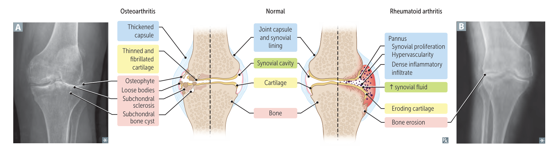
- Synovitis: Synovial lining becomes inflamed and hypertrophied.
- Pannus Formation: The inflamed synovium transforms into an aggressive a pannus (a mass of synovium, inflammatory cells, and granulation tissue).
- Erosion: The pannus invades and destroys adjacent cartilage (via MMPs) and bone (via RANKL-mediated osteoclast activation).
Clinical features
- Articular (Joints):
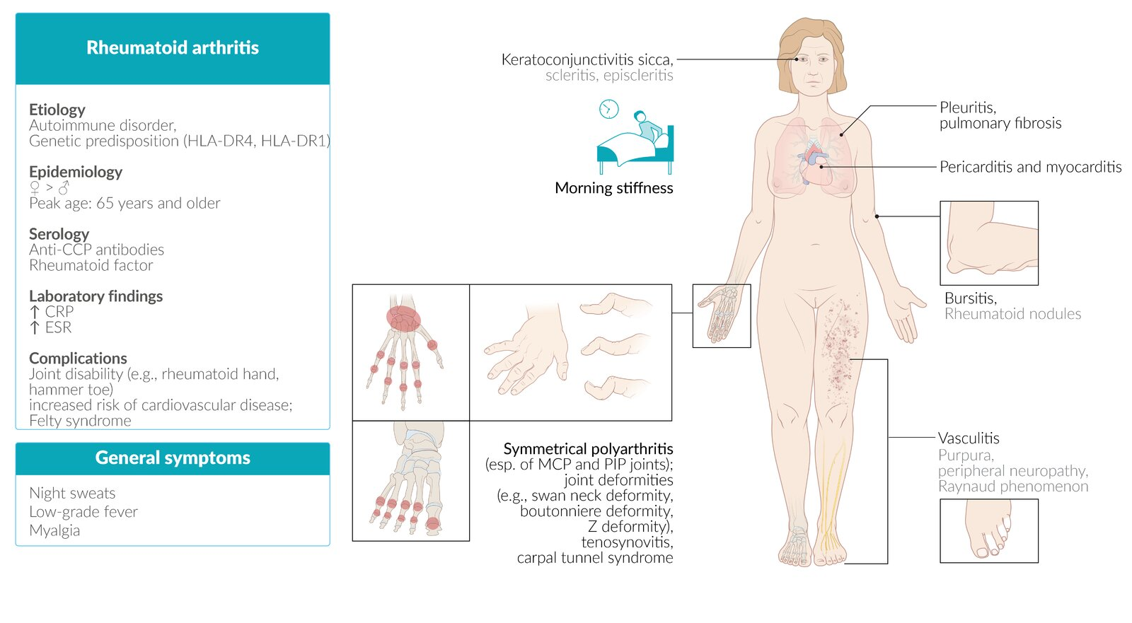
- Symmetric polyarthritis: Hallmark feature.
- Joints involved: Typically affects small joints of the hands and feet, such as the metacarpophalangeal (MCP), proximal interphalangeal (PIP), and metatarsophalangeal (MTP) joints, as well as the wrists.
- Morning stiffness: Lasts >1 hour and improves with activity (unlike osteoarthritis).
- Spared joints: The distal interphalangeal (DIP) joints are typically spared.
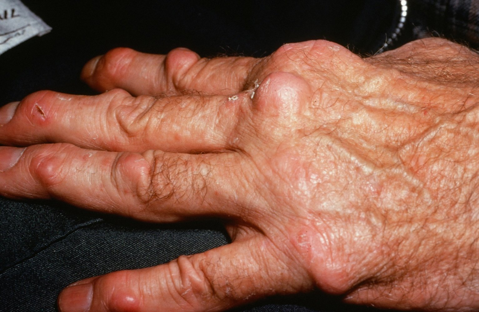
- Deformities (late-stage): Ulnar deviation of fingers, swan-neck deformity (PIP hyperextension, DIP flexion), and Boutonnière deformity (PIP flexion, DIP hyperextension).
- Extra-articular Manifestations:
- Constitutional: Fever, fatigue, weight loss, and malaise are common.
- Rheumatoid Nodules: Most common extra-articular finding; firm, subcutaneous nodules over extensor surfaces or pressure points.
- Pulmonary: Interstitial lung disease, pleuritis, and pulmonary nodules.
- Cardiovascular: Increased risk of premature atherosclerosis and coronary artery disease; pericarditis.
- Hematologic: Anemia of chronic disease, thrombocytosis.
- Ocular: Scleritis, episcleritis, and secondary Sjögren’s syndrome.
Subtypes and variants
Atlantoaxial subluxation (Vertebral subluxation)
- Definition: a potentially life-threatening complication caused by the inflammatory destruction of the ligaments affecting the atlantoaxial joint and the intervertebral joints
- Clinical features
- Pain and stiffness of the neck (typically early-morning neck pain at rest)
- Head tilt
- Neurological deficits
- Cervical radiculopathy with peripheral paresthesias of the upper limb c
- In some cases, symptoms of high spinal cord compression
- Slowly progressive spastic quadriparesis
- Hyperreflexia or positive Babinski reflex
- Respiratory insufficiency
- Diagnostics
- Extension and flexion x-rays of the cervical spine
- MRI
Warning
Endotracheal intubation can acutely worsen the subluxation and cause compression of the spinal cord and/or vertebral arteries.
Diagnostics
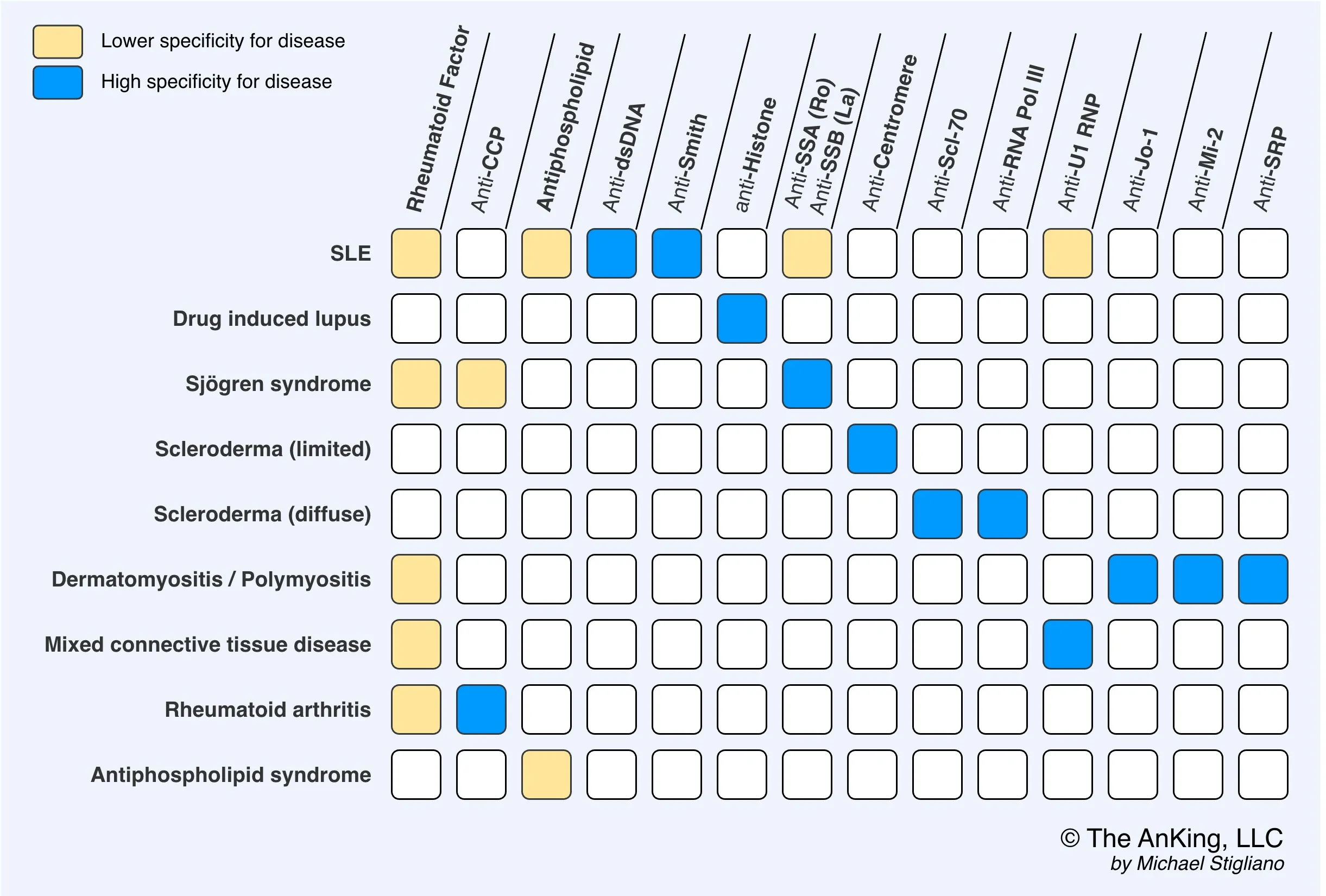
- Anticitrullinated peptide antibodies (ACPA), e.g., anticyclic citrullinated peptide (anti-CCP)
- Tissue inflammation causes arginine residues in proteins such as vimentin to be enzymatically converted into citrulline through a process called citrullination. This alters the shape of the proteins, which can then serve as neoantigens that generate an immune response.
- Rheumatoid factor (RF): IgM autoantibodies against the Fc region of IgG antibodies t
Differential diagnostics
- Parvovirus B19 will presents with similar symptoms, except for rash, normal ESR, and acute onset
- See Osteoarthritis > Differential diagnosis
Osteoarthritis (OA) vs Rheumatoid Arthritis (RA)
Feature Osteoarthritis (OA) Rheumatoid Arthritis (RA) Mechanism Mechanical “Wear & Tear” Autoimmune (Pannus) Stiffness < 30 min (Worse w/ use) > 1 hr (Better w/ use) Symmetry Asymmetric Symmetric Key Joints DIP (Heberden), PIP, Knees MCP, PIP, Wrist (Spares DIP) Labs Normal +Anti-CCP (Specific), +RF, ↑ ESR X-ray Osteophytes, Sclerosis Marginal Erosions, Osteopenia Treatment NSAIDs, Acetaminophen DMARDs (Methotrexate) 1. Stiffness
- OA (<30m): “Gelling.” Synovial fluid gets thick at rest. Movement quickly warms and lubricates it.
- RA (>1h): Edema. Inflammatory fluid pools during sleep. Takes time to mechanically pump/drain the boggy joint.
2. Pain
- OA (Worse w/ use): Mechanical. Bone-on-bone friction compresses exposed nerve endings.
- RA (Better w/ use): Washout. Movement flushes out stagnant inflammatory cytokines and fluid, relieving pressure.
3. Pathology
Link to original
- OA (Osteophytes): Construction. Bone grows spurs to widen surface area and stabilize the failing joint.
- RA (Erosions): Destruction. Pannus (granulation tissue) releases enzymes/RANKL that eat into the bone.
Treatment
- Immediate / Symptomatic Bridge:
- NSAIDs or low-dose systemic glucocorticoids (e.g., Prednisone) for rapid symptom control while awaiting DMARD onset (taper and d/c once DMARD effective).
- First-line (All newly diagnosed moderate-to-severe RA):
- Non-biologic DMARD: Methotrexate (MTX) is initial drug of choice. c
- Mechanism: Dihydrofolate reductase inhibition.
- Co-prescription: Always administer with folic acid to reduce GI, mucosal, and hematologic toxicity.
- Baseline workup: CBC, LFTs, serum Cr, CXR (screen for pulmonary fibrosis), viral hepatitis panel, pregnancy test (contraindicated in pregnancy/teratogenic).
- Monitoring: Routine CBC and LFTs (hepatotoxicity, cytopenias).
- Alternative Non-biologic DMARDs:
- Hydroxychloroquine: For mild disease or pregnancy; requires baseline and annual retinal exams (retinopathy/bull’s eye maculopathy).
- Sulfasalazine: Risk of hemolysis in G6PD deficiency, stomatitis, oligospermia.
- Leflunomide: Alternative if MTX intolerant.
- Non-biologic DMARD: Methotrexate (MTX) is initial drug of choice. c
- Second-line / Inadequate response after 3–6 months of MTX:
- Biologic DMARDs:
- TNF-alpha inhibitors: Infliximab, Adalimumab, Etanercept, Golimumab, Certolizumab.
- Non-TNF biologics: Rituximab (anti-CD20), Tocilizumab (anti-IL-6), Abatacept (CTLA-4-Ig).
- Pre-biologic screening: Must perform PPD/IGRA (latent TB) and Hepatitis B/C testing prior to initiation.
- Rule: Never combine two biologic DMARDs due to severe immunosuppression/infection risk.
- Biologic DMARDs:
- Refractory Disease:
- Targeted Synthetic DMARDs: JAK inhibitors (e.g., Tofacitinib, Baricitinib).
- Surgical Intervention: Total joint arthroplasty for end-stage joint destruction.
Complications
- Amyloidosis > AA amyloidosis (secondary amyloidosis) c
- Septic arthritis