Etiology
- Risk Factors
- Age: Most significant risk factor.
- Obesity: Increases mechanical stress on weight-bearing joints (knees, hips).
- Female gender: More common in women, especially post-menopause.
- Joint Trauma/Overuse: Previous injuries or occupations with repetitive stress can predispose to OA.
- Genetics: A family history plays a role, particularly in OA of the hand and hip.
Clinical Features
- History:
- Pain: Deep, aching; worse with activity/weight-bearing, relieved by rest (advanced disease may have pain at rest/night).
- Stiffness: Morning stiffness < 30 minutes; “gelling” phenomenon (transient stiffness after brief inactivity).
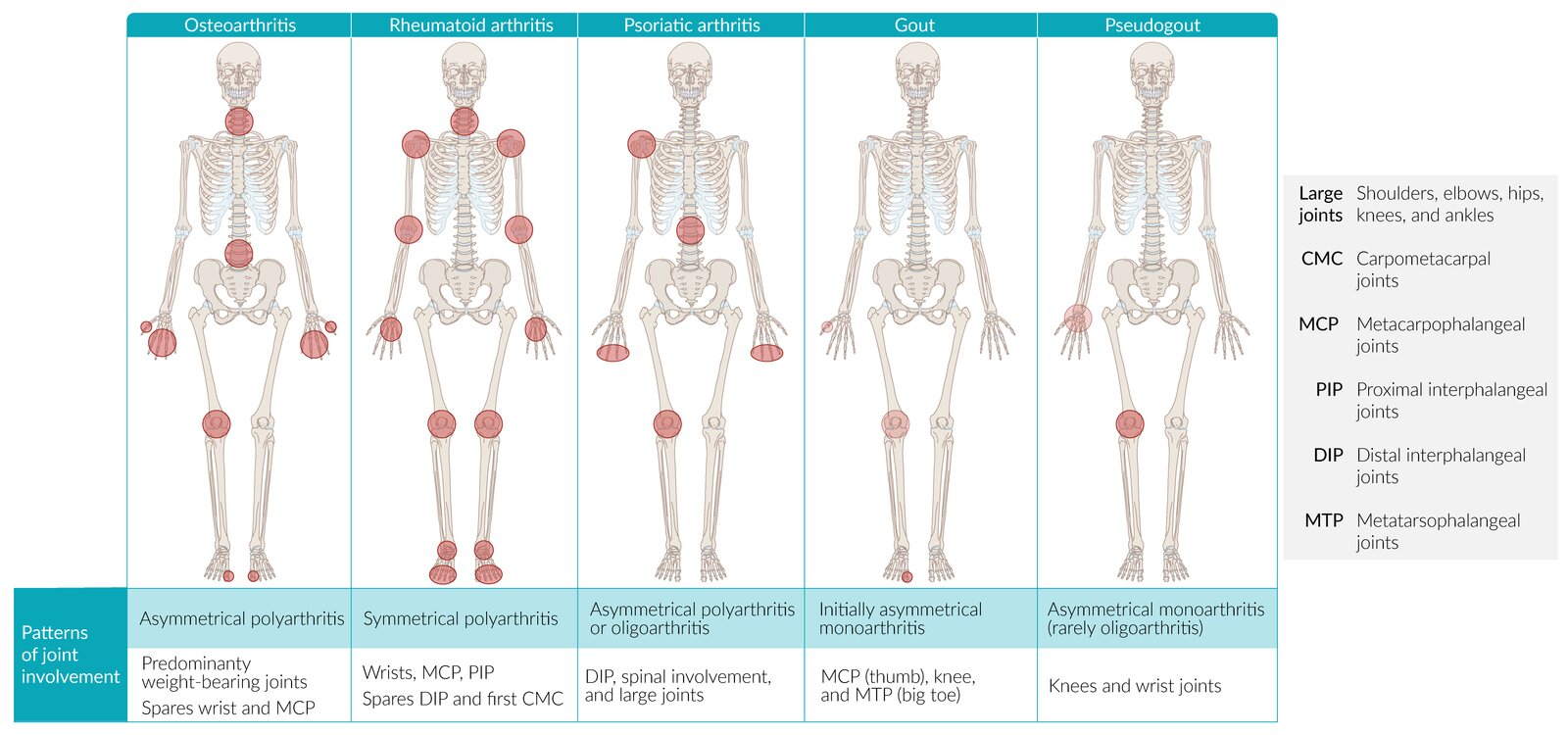
- Distribution: Asymmetric or symmetric; weight-bearing joints (knees, hips), spine (cervical/lumbar), and hands.
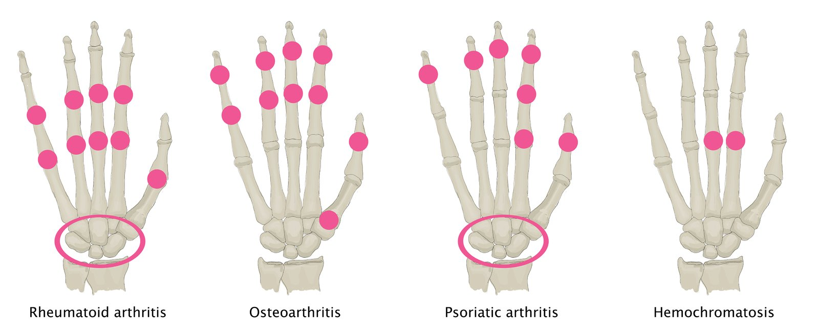
- Hand involvement: DIP joints and PIP joints, 1st CMC joint (spares MCP joints and wrists).
- Absence of constitutional symptoms (no fever, weight loss, or fatigue).
- Physical Examination:
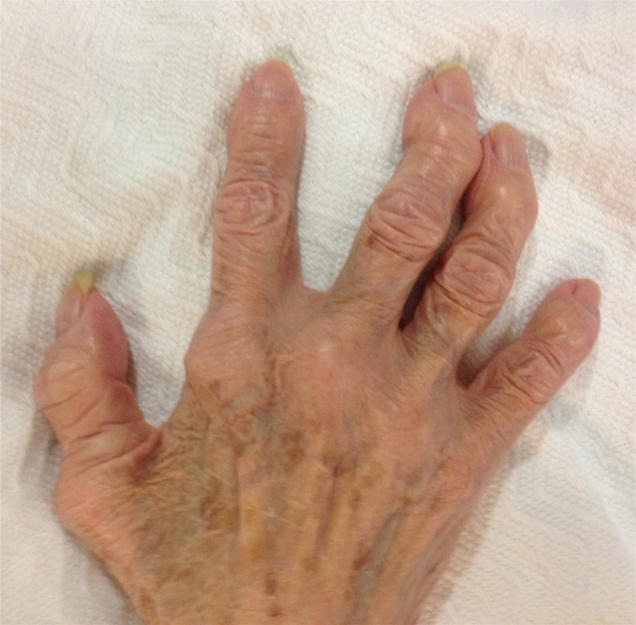
- Bony enlargement & joint line tenderness.
- Crepitus on active/passive ROM.
- Decreased active and passive ROM.
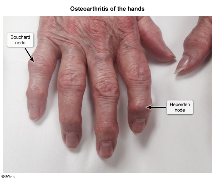
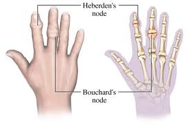
- Heberden nodes (osteophytes at DIP joints).
- Bouchard nodes (osteophytes at PIP joints).
- “Squaring” of the hand (1st CMC joint osteophytes/subluxation).
- Minimal/cool joint effusion without prominent warmth or erythema.
- Bony enlargement & joint line tenderness.
Diagnosis
- Initial / Best Initial Test: Clinical diagnosis (often sufficient based on age, typical hx, and PE findings).
- Imaging:
- Plain Radiographs (X-ray): Weight-bearing views (knee/hip).
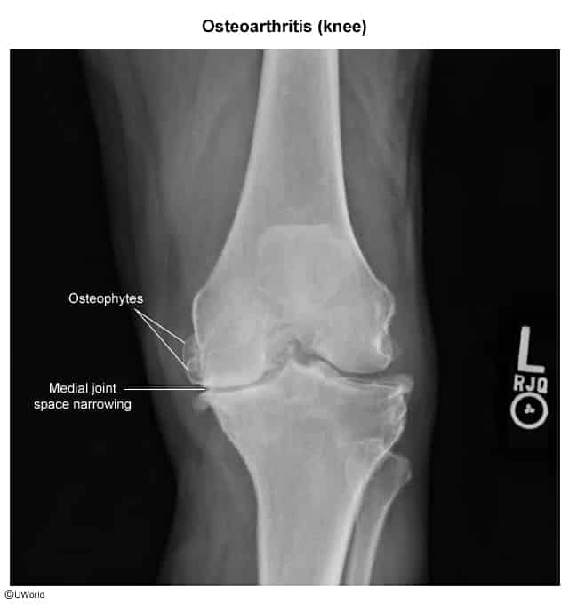
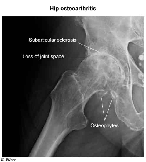
- Classic 4 Radiographic Findings:
- Asymmetric joint space narrowing (loss of articular cartilage).
- Subchondral sclerosis (bone thickening/hardening).
- Osteophyte formation (bony spurs at margins).
- Subchondral cysts (geodes).
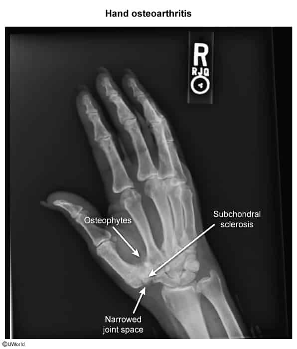
- Key Labs:
- ESR / CRP: Normal (rules out systemic inflammatory arthritides).
- Synovial Fluid Analysis (Arthrocentesis):
- Indicated only if diagnosis is uncertain or to exclude infection/crystals during an acute effusion.
- Findings: Non-inflammatory (clear/yellow, WBC < 2,000/mm³, < 25% PMNs, negative Gram stain/crystals).
Differential diagnosis
- Traditionally, osteoarthritis (OA) has been classified as a non-inflammatory or “wear-and-tear” arthritis
- The inflammation is generally less pronounced
- It doesn’t typically feature systemic inflammation markers
- It doesn’t involve autoimmune mechanisms as its primary cause
- Morning stiffness is typically shorter in duration
Osteoarthritis (OA) vs Rheumatoid Arthritis (RA)
Feature Osteoarthritis (OA) Rheumatoid Arthritis (RA) Mechanism Mechanical “Wear & Tear” Autoimmune (Pannus) Stiffness < 30 min (Worse w/ use) > 1 hr (Better w/ use) Symmetry Asymmetric Symmetric Key Joints DIP (Heberden), PIP, Knees MCP, PIP, Wrist (Spares DIP) Labs Normal +Anti-CCP (Specific), +RF, ↑ ESR X-ray Osteophytes, Sclerosis Marginal Erosions, Osteopenia Treatment NSAIDs, Acetaminophen DMARDs (Methotrexate) 1. Stiffness
- OA (<30m): “Gelling.” Synovial fluid gets thick at rest. Movement quickly warms and lubricates it.
- RA (>1h): Edema. Inflammatory fluid pools during sleep. Takes time to mechanically pump/drain the boggy joint.
2. Pain
- OA (Worse w/ use): Mechanical. Bone-on-bone friction compresses exposed nerve endings.
- RA (Better w/ use): Washout. Movement flushes out stagnant inflammatory cytokines and fluid, relieving pressure.
3. Pathology
Link to original
- OA (Osteophytes): Construction. Bone grows spurs to widen surface area and stabilize the failing joint.
- RA (Erosions): Destruction. Pannus (granulation tissue) releases enzymes/RANKL that eat into the bone.
Management
- First-line (Non-Pharmacologic - Foundational for all patients):
- Weight loss (reduces mechanical load, particularly for knee/hip OA).
- Physical therapy / low-impact exercise (e.g., quadriceps strengthening, swimming, cycling). c
- Biomechanical interventions: Contralateral cane use, lateral wedge insoles, shock-absorbing footwear.
- First-line (Pharmacologic - Symptom Relief):
- Topical NSAIDs (e.g., topical diclofenac): Preferred initial medication, especially for knee OA and elderly patients (minimal systemic SEs). c
- Oral NSAIDs (e.g., celecoxib, naproxen, ibuprofen): Most effective oral pharmacotherapy. Use lowest effective dose; add PPI in patients at high risk for GI bleeding. Monitor renal function and BP/CV risk.
- Note: Acetaminophen has limited efficacy compared to NSAIDs; topical capsaicin is a secondary adjuvant.
- Second-line / Refractory to First-Line Oral/Topical Agents:
- Intra-articular (IA) corticosteroid injections: Short-term relief (weeks to months) for acute moderate-to-severe exacerbations/effusions.
- Duloxetine: SNRI effective as adjunctive therapy for chronic OA pain (especially with comorbid depression or central sensitization).
- Refractory / Advanced Disease (Definitive):
- Total Joint Arthroplasty (TJA) (e.g., total knee/hip replacement).
- Indication: Severe pain, refractory to conservative therapy, leading to significant impairment in activities of daily living (ADLs) with radiographic evidence of advanced disease.
Tip
Pharmacotherapy should only be used as a short-term treatment in symptomatic patients; long-term therapy is associated with many adverse effects.