Proximal injury: supracondylar fracture of humerus
Distal injury
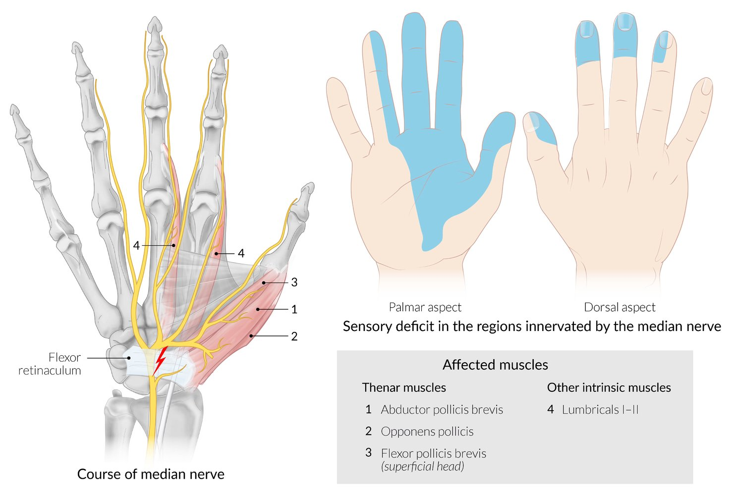
Carpal tunnel compression
Wrist laceration (suicide attempt)
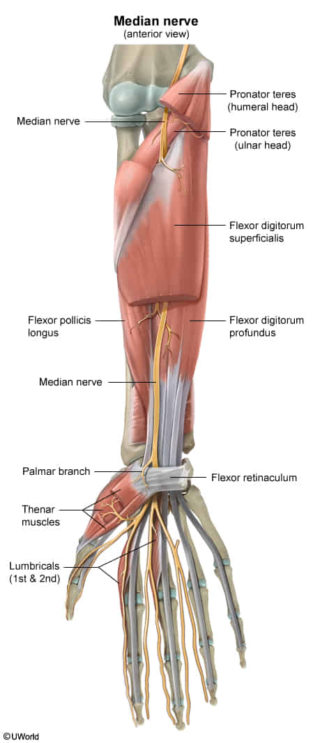
Pronator teres syndrome: compression of the median nerve between the humeral and ulnar heads of the pronator teres
Clinical features
Carpal Tunnel Syndrome (Distal Compression):
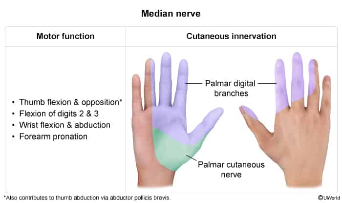
Sensory: Numbness, paresthesias, and pain in the palmar aspect of the first 3.5 digits (thumb, index, middle, and radial half of ring finger).
Nocturnal Symptoms: Symptoms characteristically awaken patient from sleep; classic “flick sign” (shaking hand relieves pain).
Motor: Weakness of thumb abduction (abductor pollicis brevis) and opposition (opponens pollicis); advanced cases present with thenar atrophy.
Anatomic Pearl: Sensation over the thenar eminence is spared in CTS because the palmar cutaneous branch arises proximal to the carpal tunnel and passes superficial to the transverse carpal ligament.
Provocative Tests:
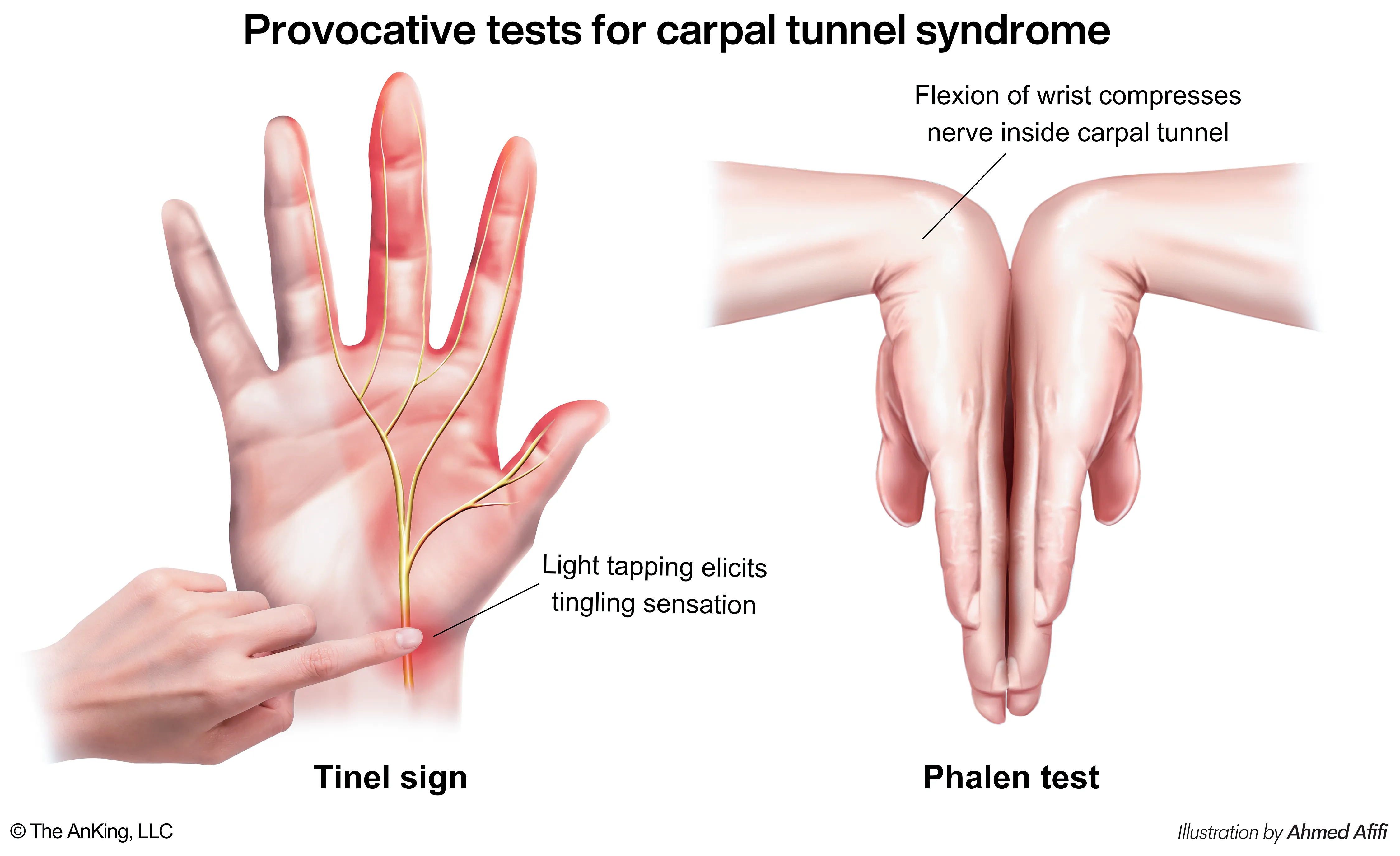
Phalen Test: Paresthesias reproduced by 60 seconds of maximal unforced wrist flexion.
Tinel Sign: Percussion over volar wrist reproduces paresthesias in median nerve distribution.
Durkan Test (Carpal Compression): Direct pressure over carpal tunnel for 30 seconds (highest sensitivity and specificity).
Proximal Median Nerve Lesions (Pronator Teres Syndrome / Elbow Trauma):
Sensation over the thenar eminence is impaired (involves palmar cutaneous branch). c
Weakness of wrist flexors, forearm pronators, and finger flexors.
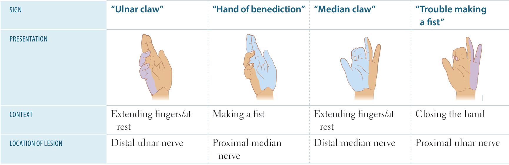
“Hand of benediction”: Inability to flex digits 1–3 when attempting to make a fist.
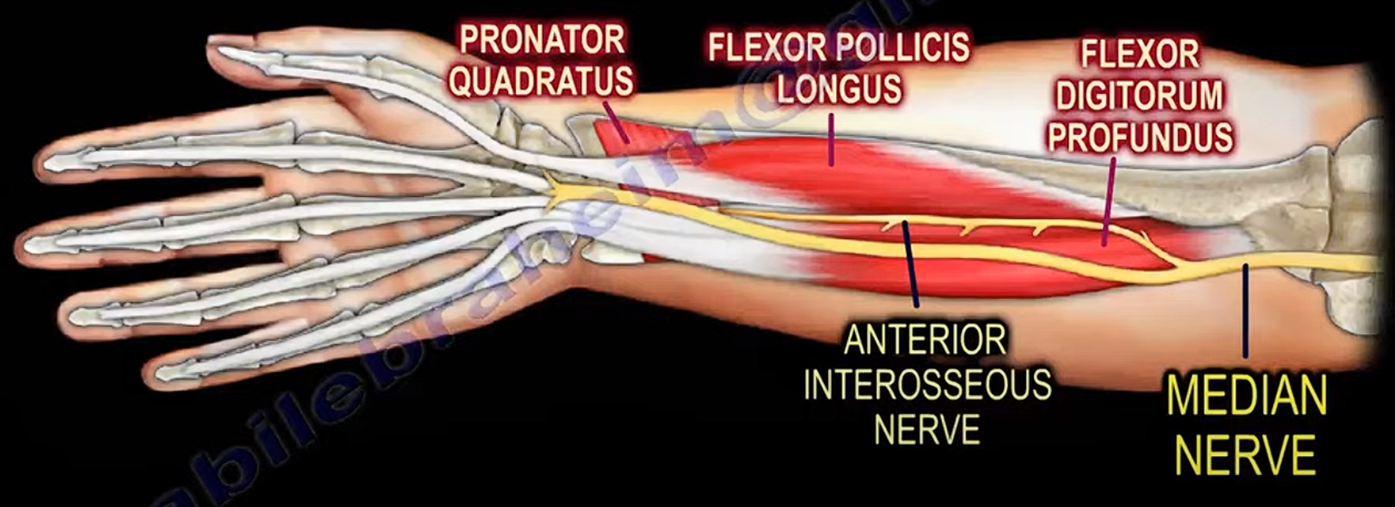
Anterior Interosseous Nerve (AIN) Syndrome:
Pure motor deficit (no sensory loss).
Inability to make the “OK” sign (impairment of flexor pollicis longus and flexor digitorum profundus to the index finger).
Motor deficits
The key is whether flexor digitorum profundus is damaged.
Nerves controlling flexor digitorum profundus branches out below the elbow. So it’s spared in distal lesion, but included in proximal lesion. So the claw hand is more prominent in distal lesion.
Anterior Interosseous Nerve -> flexor digitorum profundus -> flexion of index and middle finger
Flexor digitorum profundus
has dual innervation: the medial part is supplied by ulnar nerve
Controls wrist, MCP, and DIP joints: flexion
Flexor digitorum superficialis
Controls wrist, MCP, and PIP joints: flexion
Lumbricals and interossei
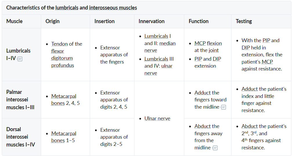
Ape hand: inability to oppose and abduct the thumb due to injury of the proximal or distal median nerves impairing the thenar muscles’ functions
Carpal tunnel syndrome
Results from a lesion that reduces the size of the carpal tunnel (fluid retention, infection, dislocation of lunate bone)
Median nerve – most sensitive structure in the carpal tunnel and is the most affected
Risk factors
Manual work: increased risk in workers using vibrating tools or prolonged, forceful, and repetitive flexion/extension of the wrist
Cccumulation of glycosaminoglycans/mucin causes myxedematous soft-tissue swelling within the carpal tunnel
Clinical manifestations:
History:
Numbness, tingling, and paresthesias in the median nerve distribution (palmar aspect of thumb, index, middle finger, and radial half of ring finger).
Symptoms worse at night; pts wake up and shake hand for relief (“flick sign”).
Clumsiness, dropping objects, difficulty buttoning shirts or opening jars.
Physical Exam:
Provocative Maneuvers:
Phalen test: Palmar paresthesias reproduced by maximum wrist flexion held for 60 seconds.
Tinel sign: Light tapping over the volar carpal tunnel reproduces paresthesias.
Durkan test (Carpal Compression Test): Direct pressure over carpal tunnel reproduces symptoms (most sensitive/specific physical test).
Motor Deficits (Late/Severe): Weakness in thumb abduction (abductor pollicis brevis) and opposition (opponens pollicis); thenar muscle atrophy.
Sensory Sparing: Sensation over the thenar eminence is preserved (palmar cutaneous branch arises proximal to and passes superficial to the carpal tunnel).
Diagnostics
Nerve Conduction Studies (NCS):Gold standard for confirmation. Shows ↓ conduction velocity and ↑ latency across the wrist.
Management
First-line (Mild-to-Moderate Symptoms / No Atrophy):
Nocturnal wrist splinting in a neutral position (prevents wrist flexion during sleep). c
Activity modification and ergonomic adjustments.
Treat underlying secondary etiology (e.g., levothyroxine for hypothyroidism; conservative treatment for pregnancy).
Second-line (Refractory to Splinting / Moderate Symptoms):
Local corticosteroid injection into the carpal tunnel (provides short-to-medium-term symptom relief).
Note: Oral NSAIDs and oral steroids show limited efficacy compared to splinting/injections.