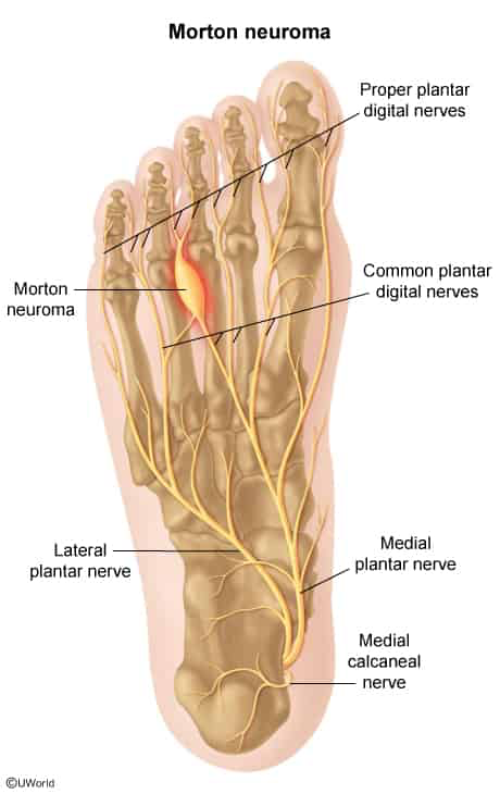
Plantar forefoot pain (burning, sharp, or cramping) radiating to adjacent toes.
Paresthesias or numbness in the interdigital space.
Sensation of “walking on a marble” or a “folded sock” in the shoe.
Symptoms worsen with weight-bearing or tight shoes; relieved by rest, removing shoes, and foot massage.
Physical Examination:
Mulder Sign (Mulder Click): Palpable click and reproduction of pain when compressing the medial and lateral metatarsal heads simultaneously with one hand while applying dorsal-plantar pressure to the interspace with the other. c
Point tenderness localized strictly to the plantar intermetatarsal space.
No significant erythema, warmth, or dorsal joint swelling.
Diagnosis
Initial / Screening & Gold Standard: Primarily a clinical diagnosis based on characteristic Hx and (+) Mulder sign.
Imaging (reserved for atypical presentations or refractory cases):
Ultrasound (US): Diagnostic imaging of choice if uncertain; demonstrates a hypoechoic, well-defined mass in the intermetatarsal space.
MRI: High sensitivity and specificity; reserved for pre-op planning or ambiguous cases.
Differential Diagnostics
Metatarsal Stress Fracture:
Diff: Pain and point tenderness on the dorsal metatarsal shaft (not interdigital webspace); (+) pain with direct bone tapping/axial loading; history of abrupt increase in training; XR shows periosteal reaction/callus (or MRI/bone scan if acute).
Tarsal Tunnel Syndrome:
Diff: Entrapment of the posterior tibial nerve behind the medial malleolus; pain/paresthesias involve the entire plantar aspect of the foot/sole; (+) Tinel sign posterior to the medial malleolus.
Plantar Fasciitis:
Diff: Pain localized to the plantar-medial calcaneus/heel; maximal with the first steps in the morning or after prolonged rest.
Freiberg Disease:
Diff: Avascular necrosis of the 2nd metatarsal head; typically seen in adolescent females; localized swelling and pain directly over the dorsal 2nd MTP joint; XR shows flattening and sclerosis of the metatarsal head.