Epidemiology
Etiology
- Population: Military recruits, competitive runners, ballet dancers, gymnasts.
- Mechanisms & Triggers:
- Abrupt ↑ in training volume, frequency, or intensity without adequate recovery.
- Repetitive mechanical overload exceeding bone remodeling capacity.
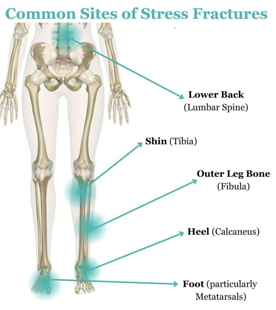
- Common Sites:
- 2nd and 3rd metatarsal shafts (most common overall; “march fracture”).
- Posteromedial tibial shaft (most common lower leg). c
- Fibula, calcaneus, navicular, femoral neck.
- Risk Factors:
- Female Athlete Triad: Low energy availability/eating disorder, amenorrhea/oligomenorrhea, ↓ BMD (osteopenia/osteoporosis).
- Nutritional deficiencies: Low Vit D, inadequate Ca intake, anorexia nervosa
- Consider these if happens in male c
- Biomechanical abnormalities: Cavus feet (high arch), leg-length discrepancy.
Tip
The female athlete triad syndrome is associated with an increased risk of stress fractures.
Pathophysiology
Clinical features
- History:
- Insidious onset of localized, aching pain.
- Initially occurs only with weight-bearing or end of training session; relieved by rest.
- Progresses to pain during daily ambulation, and eventually rest/night pain.
- Physical Exam:
- Focal point tenderness directly over the affected bone cortex (hallmark). c
- Localized soft tissue edema and erythema.
- Positive “tuning fork” or percussion test (vibration reproduces sharp, localized pain).
- Antalgic gait.
Diagnostics
- X-ray: recommended initial imaging study
- Radiographic features of stress fractures include:
- Fracture line: line of faint lucency
- Indirect features: periosteal thickening, increased cortical density, formation of a callus, endosteal thickening and/or sclerosis
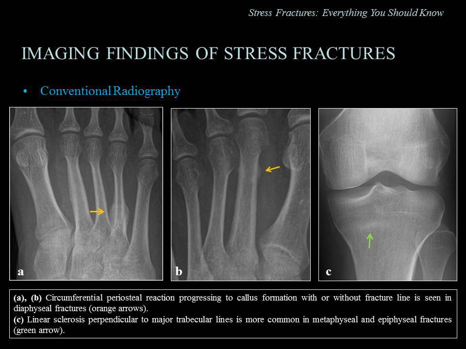
- Often normal in the first 2–3 weeks of disease onset
- Radiographic features of stress fractures include:
Differential Diagnostics
- Medial Tibial Stress Syndrome (“Shin Splints”):
- Diff by diffuse tenderness along posteromedial tibial border (>5 cm) without focal point tenderness; XR normal; MRI shows periosteal edema without cortical line or deep marrow edema.
Treatment
<% tp.file.cursor() %>