Epidemiology
Etiology
Strong association with Epstein-Barr virus (EBV) c
Immunodeficiency: e.g., organ or cell transplantation, immunosuppressants, HIV infection , chemotherapy
Autoimmune diseases (e.g., rheumatoid arthritis , sarcoidosis )
Hodgkin lymphoma vs non-Hodgkin lymphoma
Feature Hodgkin Lymphoma (HL )Non-Hodgkin Lymphoma (NHL )Key Cell Reed-Sternberg (“owl-eye”)Malignant lymphocytes (B or T cell) Spread Contiguous (orderly, predictable)Non-contiguous (disseminated)Presentation Localized, single node group (e.g., cervical, mediastinal ) Multiple peripheral nodes, extranodal common ”B” Symptoms Common (fever, night sweats, wt loss)Less common Age Bimodal (20s, >50s)Typically >50 years old Prognosis High cure rate Variable; depends on subtype Key Associations EBV EBV HTLV-1 , HHV -8
Link to original
Pathophysiology
Clinical features
Painless Lymphadenopathy (LAD) :
Firm, rubbery, mobile or matted, non-tender nodes.
Anatomic distribution: Cervical (60–70%) > Supraclavicular > Axillary > Mediastinal.
Contiguous spread via lymphatic channels.
B Symptoms (correlate with advanced disease and poorer prognosis):
Unexplained fever (>38°C / 100.4°F).
Drenching night sweats.
Unintentional weight loss (>10% of body weight over 6 mo).
Pathognomonic / High-Yield Signs :
Alcohol-induced pain : Immediate severe nodal pain within minutes of alcohol consumption.Pel-Ebstein fevers : Alternating cycles of high fever for 1–2 weeks followed by afebrile periods (rare but classic).Generalized pruritus : Unexplained, severe itching without a primary rash.
Mass Effect (Mediastinal Mass) :
Non-productive cough, dyspnea, retrosternal chest fullness.
Superior Vena Cava (SVC) Syndrome : Facial plethora, JVD, upper extremity edema.
Diagnostics
Initial / Definitive Diagnostic Step : Excisional lymph node biopsy .
Note : FNA is contraindicated/inadequate (preservation of node architecture required).
Histopathology / Confirmatory :
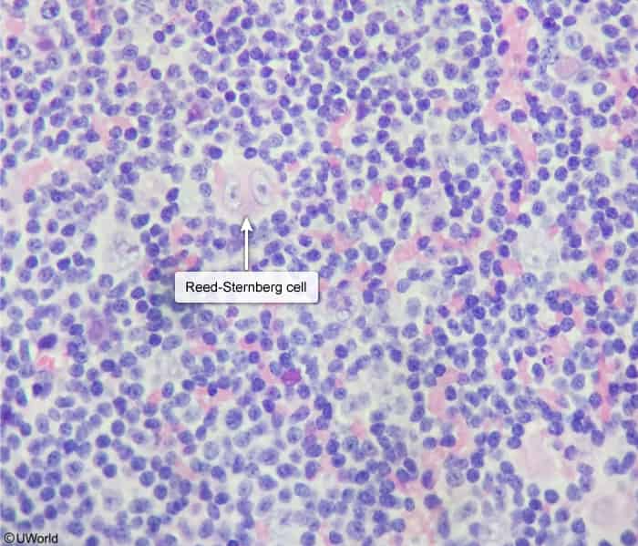
Reed-Sternberg (RS) cells : Giant, binucleated or multinucleated B-cells with prominent nucleoli (“Owl’s eye” appearance ).Immunohistochemistry (Classic HL) : CD15(+) , CD30(+) , CD20(-), CD45(-).
Staging Workup (Ann Arbor Staging) :
Whole-body FDG-PET/CT (skull base to mid-thigh) is the gold standard for staging and response assessment. c Staging Classification:
Stage I: Single lymph node region.
Stage II: ≥2 lymph node regions on the same side of the diaphragm.
Stage III: Lymph node regions on both sides of the diaphragm.
Stage IV: Extranodal involvement (e.g., bone marrow, liver, lung).
Modifier: A = No systemic symptoms; B = B symptoms present.
Key Labs & Baseline Pre-treatment Tests :
CBC: Normocytic normochromic anemia, leukocytosis, eosinophilia .
↑ ESR / CRP : Correlates with disease activity and prognosis.Serum LDH: Non-specific marker of tumor turnover.
LFTs, BUN/Cr, viral serologies (HIV, Hep B, Hep C).
Bone marrow biopsy: Only indicated if cytopenias or high-risk Stage III/IV disease with equivocal PET.
Pre-chemo Organ Function :
Echocardiogram / MUGA scan : Baseline EF prior to anthracyclines (Doxorubicin).PFTs with DLCO : Baseline pulmonary function prior to Bleomycin.
Treatment
Prognosis c
HL is highly curable; the majority of patients achieve complete remission with current multi-agent chemotherapy (ABVD) and/or radiation.
HL relapse (or recurrence): The leading cause of death for the first 8-10 years after treatment is HL relapse. Relapses primarily occur in the first 2 years, but risk remains mildly elevated for life.Second malignancy: The leading cause of death >8-10 years after treatment is second malignancy, and this risk increases with time .
Caused by radiation and chemotherapy . Transformation of healthy somatic cells into clinical solid tumors typically takes ≥8–10 years .
Complications
Late Treatment-Related Toxicities (High-Yield for Step 2 CK) :
Secondary Malignancies (leading cause of long-term mortality in cured HL survivors):
Breast Cancer : Extremely high risk in young females receiving chest/mediastinal ISRT.
Screening guideline : Annual mammogram + breast MRI starting at age 25 or 8–10 years post-radiation (whichever comes later).
Lung Cancer : Synergistic risk with cigarette smoking and chest radiation.Therapy-Related Acute Myeloid Leukemia (t-AML) / MDS : Occurs 2–5 yrs post-treatment (alkylating agents, etoposide).Thyroid Cancer : Secondary to neck radiation.
Cardiovascular Disease :
Accelerated Coronary Artery Disease (CAD), premature MI (radiation-induced endothelial injury).
Valvular heart disease (aortic/mitral stenosis or regurgitation).
Constrictive pericarditis and pericardial effusion.
Dilated cardiomyopathy (Doxorubicin toxicity; cumulative dose-dependent, permanent).
Pulmonary Toxicity :
Bleomycin-induced pulmonary fibrosis / pneumonitis : Presents with non-productive cough, dyspnea, bilateral infiltrates, decreased DLCO.Radiation pneumonitis / apical fibrosis.
Endocrine & Reproductive :
Hypothyroidism (up to 50% post-neck/mediastinal radiation; screen with annual TSH).Premature ovarian failure / Azoospermia : Infertility from alkylating agents (offer sperm/oocyte banking prior to initiation).
Disease-Related Complications :
SVC syndrome.
Tracheal compression / airway compromise.
Spinal cord compression (epidural tumor extension).
Opportunistic infections secondary to cellular immune defects (e.g., Herpes zoster , Pneumocystis jirovecii ).