Epidemiology & Risk Factors
- Primary Raynaud Phenomenon (Raynaud Disease):
- Most common (>80% of cases).
- Typically females aged 15–30 years.
- No underlying systemic disease, normal physical exam, normal nailfold capillaries, negative inflammatory markers/autoantibodies.
- Secondary Raynaud Phenomenon (Raynaud Syndrome):
- Typically onset at >30–40 years of age; equal male/female prevalence.
- Connective Tissue Diseases (CTD): Systemic Sclerosis (SSc / Scleroderma) (>90% of SSc pts), SLE, Mixed Connective Tissue Disease (MCTD), Sjögren syndrome, Dermatomyositis.
- Vascular / Occlusive: Thromboangiitis obliterans (Buerger disease), atherosclerosis, thoracic outlet syndrome (TOS).
- Occupational / Physical: Repeated vibration trauma (e.g., jackhammer, chainsaw use), frostbite.
- Medications / Drugs: Beta-blockers, ergotamines/triptans, stimulants/amphetamines, cocaine, chemotherapeutics (bleomycin, cisplatin), nicotine.
- Hematologic: Cryoglobulinemia, cold agglutinin disease, paraproteinemia.
Diagnosis
- Initial Evaluation:
- Clinical diagnosis based on history of cold/stress-induced episodic digital color changes.
- Key goal: Differentiate Primary vs. Secondary RP.
- Initial / Bedside Test:
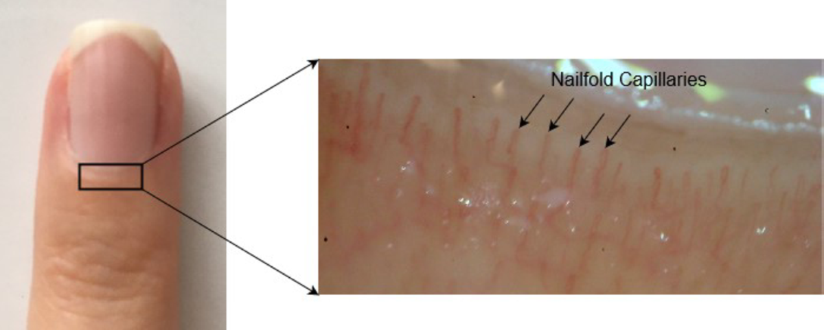
- Nailfold Capillaroscopy:
- Normal parallel, hairpin loops -> Primary RP.
- Dilated, tortuous, or dropped-out (avascular areas) loops -> Secondary RP (especially SSc/CTD).
- Key Labs (indicated if secondary RP suspected or age >30):
- Initial Screening: ANA, ESR / CRP, CBC, CMP, Urinalysis. c
- Confirmatory / Specific Autoantibodies (if ANA positive):
- Anti-Scl-70 (Anti-topoisomerase I) & Anti-Centromere: Systemic Sclerosis (diffuse vs. limited/CREST).
- Anti-dsDNA, Anti-Smith: SLE.
- Anti-U1-RNP: Mixed Connective Tissue Disease (MCTD).
- Anti-Ro/SSA, Anti-La/SSB: Sjögren syndrome.
- Cryoglobulins / Serum protein electrophoresis (SPEP): If associated with purpura, arthralgias, or renal disease.
- Vascular Imaging / Testing:
- Indicated if asymmetric presentation, absent pulses, or suspicion of proximal macrovascular occlusion.
- Arterial Duplex US or CTA / MRA / Digital Plethysmography.