Clinical Features
Acute Pulmonary :
Flu-like syndrome: Fever, chills, nonproductive cough, substernal chest pain.
Extrapulmonary hypersensitivity signs: Erythema nodosum , erythema multiforme , polyarthralgias.
Chronic Cavitary Pulmonary :
Mimics TB: Chronic productive cough, hemoptysis, night sweats, weight loss (predominantly in underlying COPD/emphysema).
Disseminated Histoplasmosis (Immunocompromised): H. capsulatum is an intracellular parasite of the monocyte-macrophage system , will disseminate via blood/lymphatics and deposit
Systemic: High fevers, severe weight loss, night sweats.
Reticuloendothelial system: Hepatosplenomegaly , generalized lymphadenopathy.
Mucocutaneous: Painful oral mucosal ulcers / tongue erosions, petechiae. c
Endocrine: Adrenal insufficiency (flank pain, hypotension, hyponatremia, hyperkalemia).
Diagnose
Initial / Screening Test :
Urine and Serum Histoplasma Antigen ELISA (rapid, highest sensitivity for acute pulmonary and disseminated disease).
Confirmatory / Gold Standard :
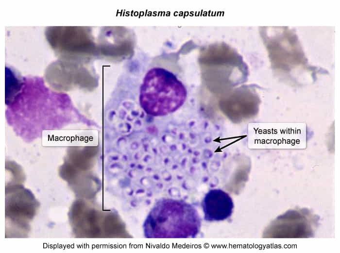
Fungal Culture (sputum, blood, bone marrow, tissue) or Histopathology .Histopathology (Wright-Giemsa or Silver stain): Small (2-5 µm) intracellular oval yeasts inside macrophages (“Histo Hides in macrophages”).
Key Labs :
Cytopenias (pancytopenia secondary to bone marrow infiltration).
↑ LFTs (alkaline phosphatase , total bilirubin).↑ LDH and ↑ Ferritin (marked elevation in severe disseminated disease).
Imaging :
CXR / Chest CT :
Acute: Patchy/reticulonodular infiltrates with hilar/mediastinal lymphadenopathy .
Chronic/Old: Buckshot calcifications (calcified pulmonary granulomas and splenic calcifications).
Chronic Cavitary: Apical cavity lesions.
Differential Diagnostics
Tuberculosis (TB)
Diff by (+) PPD / IGRA, acid-fast bacilli (AFB) on stain, cavitary lesions typically apical, no fungal antigen.
Blastomycosis :
Diff by large broad-based budding yeast , endemic to Great Lakes/Ohio River, prominent verrucous/ulcerative skin lesions and osteolytic bone lesions.
Coccidioidomycosis
Diff by large spherules containing endospores , Southwest US (San Joaquin Valley/Arizona), desert dust exposure, peripheral eosinophilia.
Sarcoidosis :
Diff by non-caseating granulomas , ↑ ACE levels , bilateral hilar lymphadenopathy without infectious etiology or fungal antigen; improves with steroids (steroids worsen untreated histoplasmosis).
Pneumocystis jirovecii Pneumonia (PJP)
Diff by diffuse bilateral ground-glass infiltrates, silver stain showing cup-shaped cysts , lack of hepatosplenomegaly or oral ulcers.