Epidemiology & Risk Factors
- Geographic Endemicity: Southwestern US (Arizona, San Joaquin Valley in California, New Mexico, West Texas), Mexico, Central/South America.
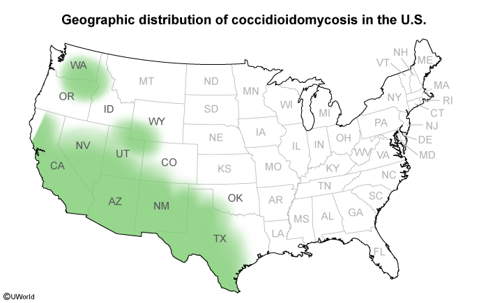
- Transmission: Inhalation of airborne arthroconidia (mold in soil/dust) after dust storms, earthquakes, construction, or farming.
- Pathology: Dimorphic fungus.
- Environment (soil, 25°C): Mold form with barrel-shaped arthroconidia.
- Tissue (human body, 37°C): Spherules filled with endospores (NOT yeast in tissue).
- Risk Factors for Disseminated Disease:
- Immunocompromised (HIV w/ CD4 < 250, transplant, anti-TNF therapy).
- 3rd trimester of pregnancy (elevated estrogen/progesterone promotes fungal growth).
- Filipino, African American, Hispanic descent.
- Advanced age, DM2.
Clinical Features
- Asymptomatic: ~60% of infected individuals.
- Acute Pulmonary Disease (“Valley Fever”):
- Symptoms: Fever, dry cough, pleuritic chest pain, fatigue, weight loss.
- “Desert Rheumatism”: Arthralgias (commonly knees and ankles).
- Cutaneous: Erythema nodosum (painful tender red shins; indicates good cell-mediated immune response) or Erythema multiforme. c
- Chronic/Progressive Pulmonary Disease:
- Persistent cough, hemoptysis, weight loss, fever, cavitary pulmonary lesions (mimics TB).
- Disseminated Disease (Extrapulmonary):
- Skin: Verrucous papules, plaques, subcutaneous abscesses.
- Musculoskeletal: Osteomyelitis, lytic bone lesions, septic arthritis.
- CNS: Subacute/chronic basilar meningitis (most severe/fatal presentation).
Diagnosis
- Initial/Screening: Serology (EIA for IgM/IgG).
- IgM (+) in 1-3 weeks; IgG (+) later (high titer IgG > 1:16 indicates risk of dissemination).
- Confirmatory/Gold Standard: Tissue Biopsy showing spherules containing endospores (H&E, GMS, or PAS stain).
- Fungal culture is diagnostic but poses a high biohazard risk to lab staff.
- Key Labs: Peripheral eosinophilia on CBC (classic USMLE clue).
- Imaging (CXR/CT):
- Ipsilateral hilar lymphadenopathy, lobar infiltrates, reticulonodular opacities, thin-walled cavities.
- Lumbar Puncture (if CNS symptoms present):
- CSF: Lymphocytic pleocytosis, low glucose, high protein, (+) CSF complement-fixing antibodies.
Differential Diagnostics
- Histoplasmosis: Diff by Mississippi/Ohio River Valleys, bird/bat guano exposure, biopsy shows intracellular small yeasts within macrophages.
- Blastomycosis: Diff by Great Lakes/Upper Midwest, broad-based budding yeasts, osteolytic bone lesions + verrucous skin lesions, lack of EN.
- Tuberculosis: Diff by positive PPD/IGRA, acid-fast bacilli (AFB) on sputum stain, upper-lobe cavitary lesions without “Desert Rheumatism”/EN triad.
- Sarcoidosis: Diff by bilateral hilar lymphadenopathy, non-caseating granulomas, ↑ ACE level, hypercalcemia, negative fungal serologies.
Management
- Uncomplicated/Mild Primary Pulmonary Disease (Immunocompetent):
- Supportive care/Observation (self-limiting in low-risk pts; follow up q3-6mo).
- Moderate-to-Severe Pulmonary OR High-Risk Patients (Immunocompromised, pregnant, DM2, severe Sx):
- First-line: Oral Fluconazole or Itraconazole for 3-6 months.
- Severe/Disseminated Disease OR Refractory Pulmonary:
- Initial: IV Liposomal Amphotericin B.
- Maintenance: Step-down to oral azole (Fluconazole/Itraconazole) for >12 months.
- Coccidioidal Meningitis:
- High-dose oral Fluconazole (lifelong therapy required to prevent relapse).
Complications
- Chronic cavitary pulmonary disease, hemoptysis, bronchopleural fistula.
- Coccidioidal Meningitis (hydrocephalus, herniation, death).
- Extrapulmonary dissemination (bone destruction, severe skin ulceration).
- High rate of disease relapse upon cessation of antifungal therapy in immunocompromised pts.