Epidemiology
Most common malignancy in patients with untreated HIV
Etiology
- Etiologic Agent: Human Herpesvirus 8 (HHV-8) / Kaposi Sarcoma-associated Herpesvirus (KSHV).
- Subtypes:
- Epidemic (AIDS-related): Most common in US; occurs with CD4 < 200/mm³ (sexually transmitted route).
- Classic (Sporadic): Elderly men of Mediterranean or Eastern European Jewish descent.
- Endemic (African): Sub-Saharan African children and young men; aggressive lymphadenopathic course.
- Iatrogenic (Post-transplant): Solid organ transplant recipients on chronic immunosuppressive therapy. c
Pathophysiology
HHV-8 infects vascular and lymphatic endothelial cells, causing them to differentiate into a mixed phenotype thought to increase oncogenic potential. The HHV-8 genome contains several viral oncogenes that inhibit cell cycle regulation and apoptosis, thereby promoting endothelial cell growth and tumorigenesis
Clinical features
- Cutaneous Lesions:
- Violaceous (purplish-red to dark brown) macules, papules, plaques, or nodules.
- Non-tender, non-pruritic, non-blanching. c
- Slit-like vascular channels, blood leaks into the surrounding interstitial dermis
- Common sites: Lower extremities, trunk, face, mucosal surfaces (hard palate, gingiva).
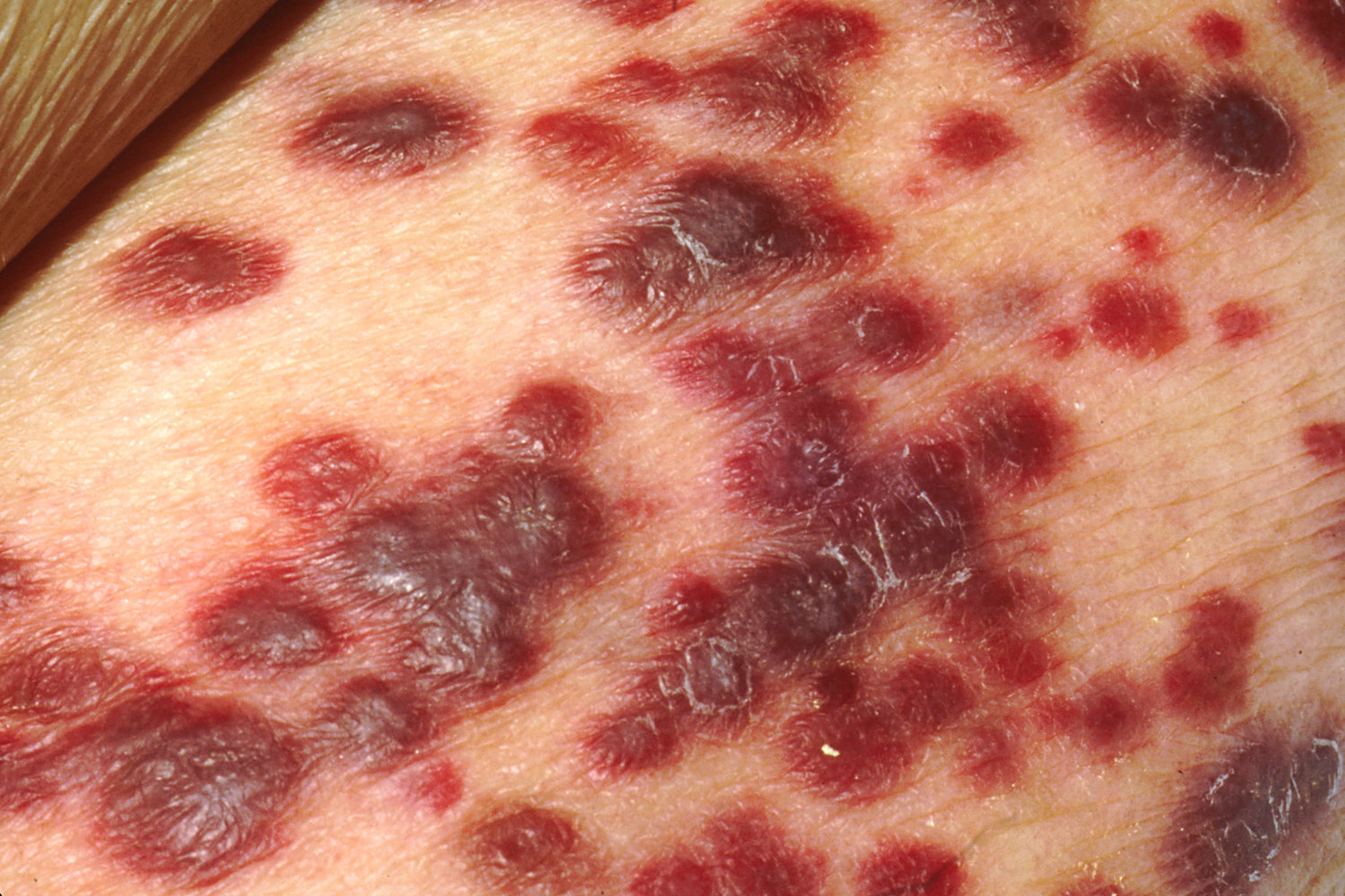
- Progression: may disseminate to the organs, particularly the gastrointestinal tract, respiratory tract, and lymph nodes.
- Gastrointestinal tract involvement: abdominal pain and diarrhea; hemorrhage and bowel obstruction in severe cases
- Often polypoid nodules
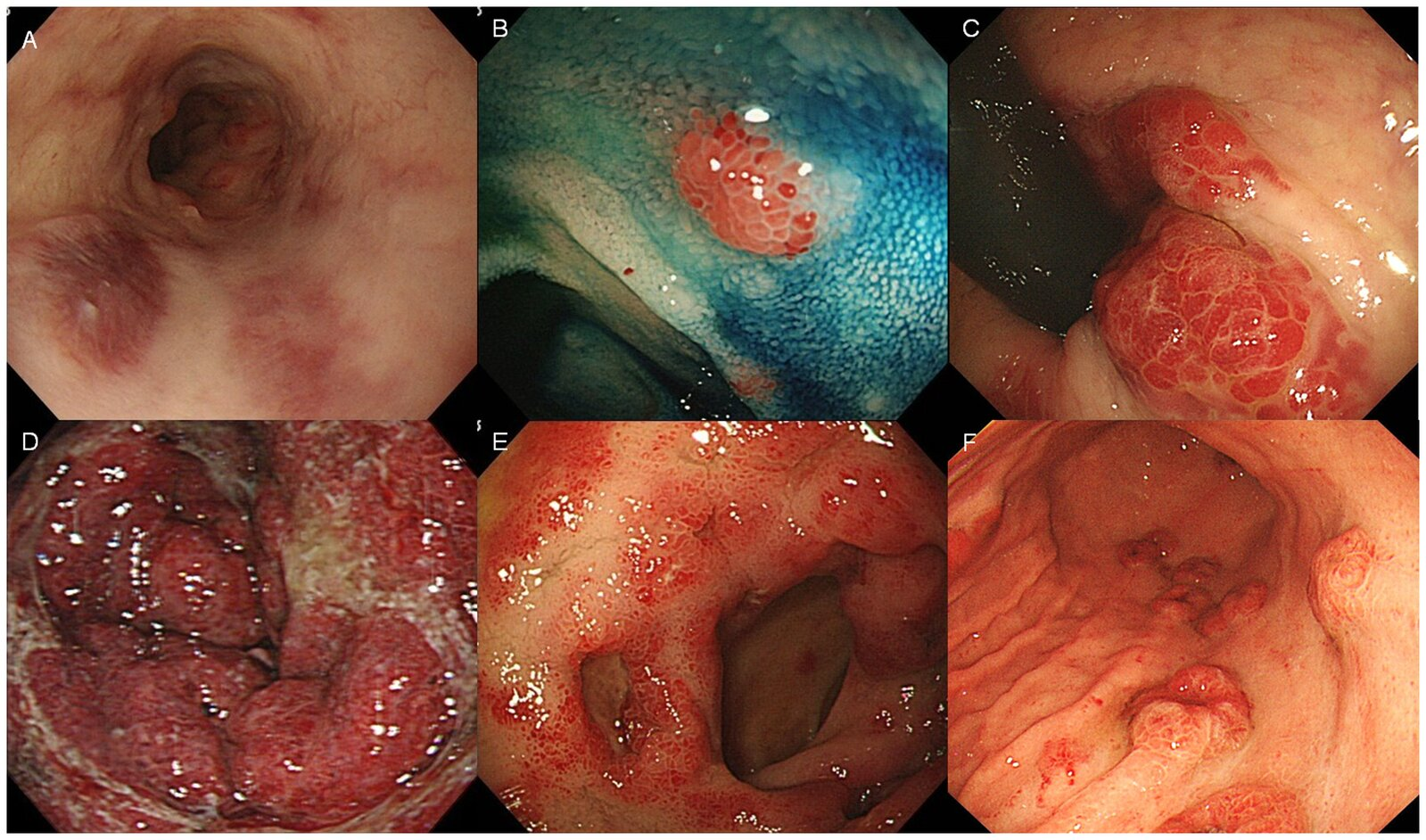
- Often polypoid nodules
- Pulmonary: Dyspnea, cough, hemoptysis, fever. CXR shows bilateral interstitial infiltrates and pleural effusions (poor prognosis).
- Gastrointestinal tract involvement: abdominal pain and diarrhea; hemorrhage and bowel obstruction in severe cases
Diagnostics
- Biopsy and histology of cutaneous or visceral lesions (indicated in all patients)
- Dermal proliferation of spindle-shaped endothelial cells with slit-like vascular spaces, red blood cell extravasation, and inflammation.
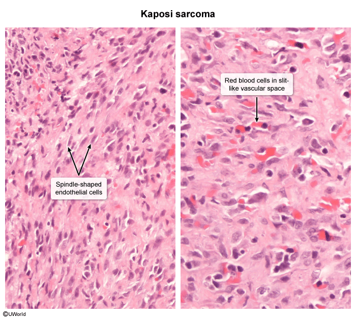
- Dermal proliferation of spindle-shaped endothelial cells with slit-like vascular spaces, red blood cell extravasation, and inflammation.
| Feature | Bacillary Angiomatosis (BA) | Kaposi Sarcoma (KS) |
|---|---|---|
| Etiology | Bartonella spp. (Bacteria) | HHV-8 (DNA Virus) |
| Histology | Capillary proliferation, plump endothelial cells | Spindle cells, slit-like vascular spaces |
| Infiltrate | Neutrophilic | Lymphocytic & Plasmacytic |
| Special Stain | Warthin-Starry stain shows organisms | N/A |
| Treatment | Antibiotics (Doxycycline, Erythromycin) | ART (first-line), Chemotherapy/Radiation for severe disease |
Differential Diagnostics
- Bacillary Angiomatosis (Bartonella henselae/quintana):
- Diff by history of cat/flea exposure; lesions are red/purple vascular papules that blanch on pressure.
- Biopsy shows neutrophilic infiltrate with Warthin-Starry silver stain (+) for bacilli (vs KS: spindle cells, lymphocytic infiltrate, HHV-8 +).
- Melanoma:
- Diff by ABCDE criteria (asymmetry, border irregularity, color variegation), lack of mucosal predilection, and S-100 / HMB-45 (+) on biopsy.
- Pyogenic Granuloma:
- Diff by solitary, rapidly growing, friable, highly vascular papule prone to profuse bleeding with minor trauma; lobular capillary proliferation on biopsy.
- Stasis Dermatitis:
- Diff by location restricted to bilateral lower extremities, association with chronic venous insufficiency/varicosities, and lack of oral/visceral lesions.
- Angiosarcoma:
- Diff by rapid aggressive growth, history of radiation therapy or chronic lymphedema (Stewart-Treves syndrome), and negative HHV-8 IHC.
Treatment
- Systemic Antiretroviral Therapy (ART):
- First-line treatment for all AIDS-related cases. Immune reconstitution often causes regression of cutaneous lesions.
- Systemic Chemotherapy:
- Indicated for extensive cutaneous disease, visceral involvement (GI/pulmonary), or progression despite ART.
- First-line agents: Liposomal doxorubicin or paclitaxel.
- Local Therapy (Symptomatic or cosmetic lesions only):
- Intralesional vinblastine, topical alitretinoin, radiation therapy, or cryotherapy.
- Transplant-related KS:
- Reduce immunosuppressive therapy (e.g., transition to mTOR inhibitors like sirolimus).