Epidemiology
Etiology
Risk factors
- For contracting HPV infection
- Environmental risk factors
- Cigarette smoking and/or exposure to second-hand smoke (for squamous cell cancer types only)
- In-utero exposure to diethylstilbestrol (DES)
Pathophysiology
Overview of terminology
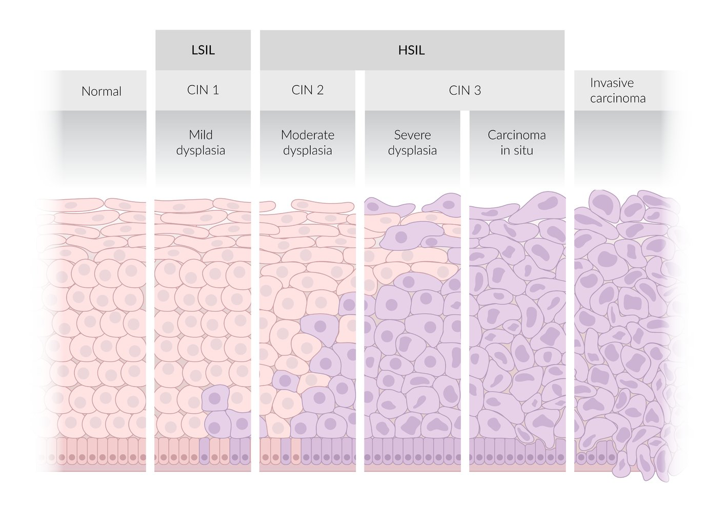
Two-tiered terminology (Bethesda system)
- High-grade squamous intraepithelial lesion (HSIL)
- Lesions with moderate to severe epithelial dysplasia that are most likely associated with persistent HPV infection
- HSIL lesions can progress to invasive cervical cancer (precancerous lesion).
- Low-grade squamous intraepithelial lesion (LSIL)
- Lesions with mild epithelial dysplasia that are typically associated with transient HPV infections
- Correlates with CIN1
Three-tiered terminology
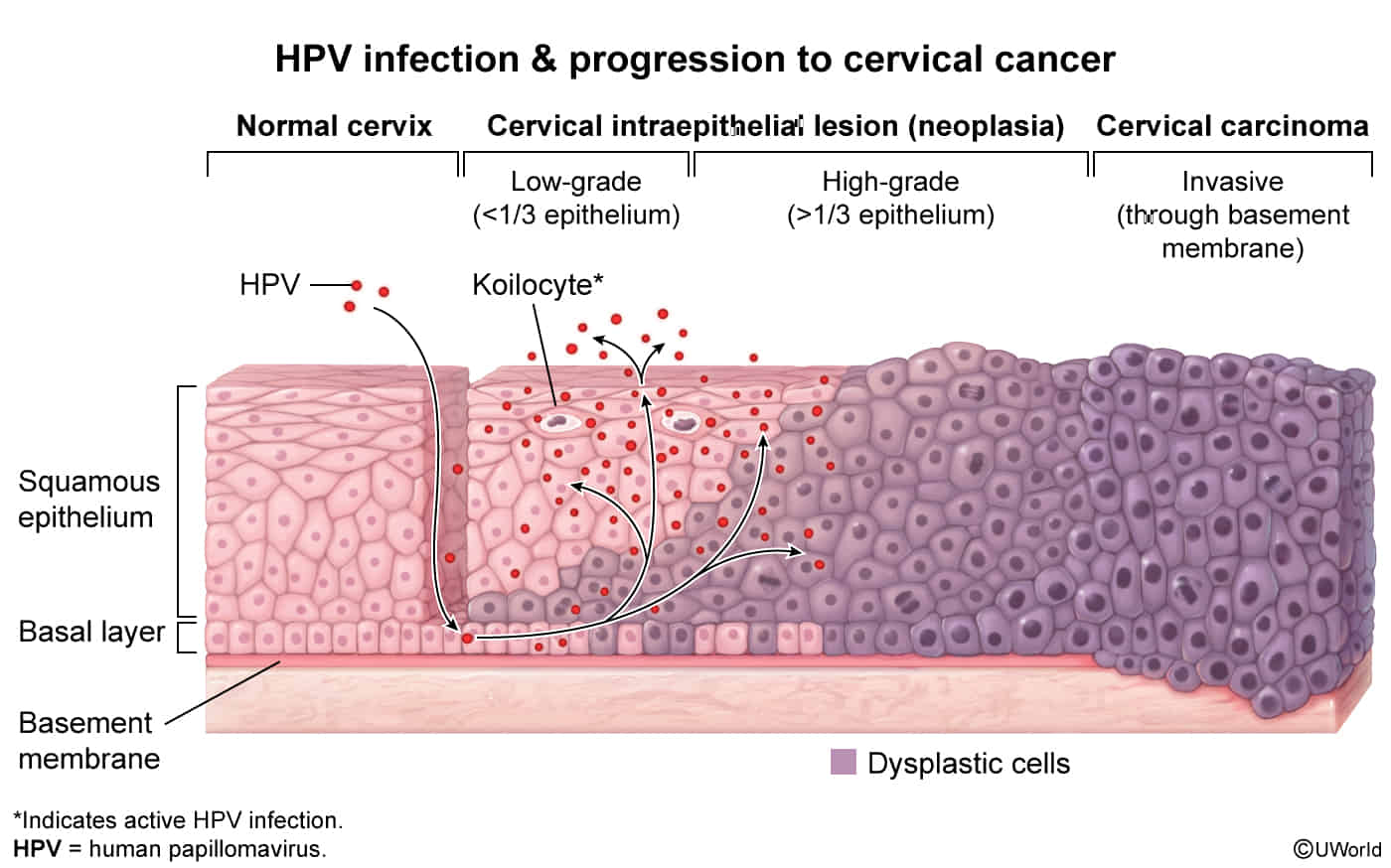
- Cervical intraepithelial neoplasia (CIN) is characterized by epithelial dysplasia that begins at the basal layer of the squamocolumnar junction and extends outward.
- Based on the degree of dysplasia, lesions are classified as CIN1 (mild), CIN2 (moderate), or CIN3 (severe).
Current standard
The current standard for reporting cervical histopathology and cytopathology to ensure optimal management is a combination of the two-tier and three-tier system (i.e., the two-tiered nomenclature with an additional qualifier using the intraepithelial neoplasia nomenclature).
- LSIL correlates with CIN1.
- HSIL correlates with CIN3 and CIN2.
- HSIL should be specified as HSIL (CIN2), HSIL (CIN3), or HSIL unspecified.
Normal cervix
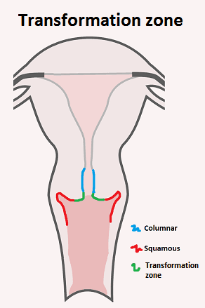
- Ectocervix: Lined by non-keratinized stratified squamous epithelium, continuous with the vaginal epithelium.
- Endocervix: Lined by simple columnar epithelium, which is mucus-secreting.
- Squamocolumnar Junction (SCJ): The sharp boundary where the squamous and columnar epithelia meet.
- Transformation Zone (TZ):
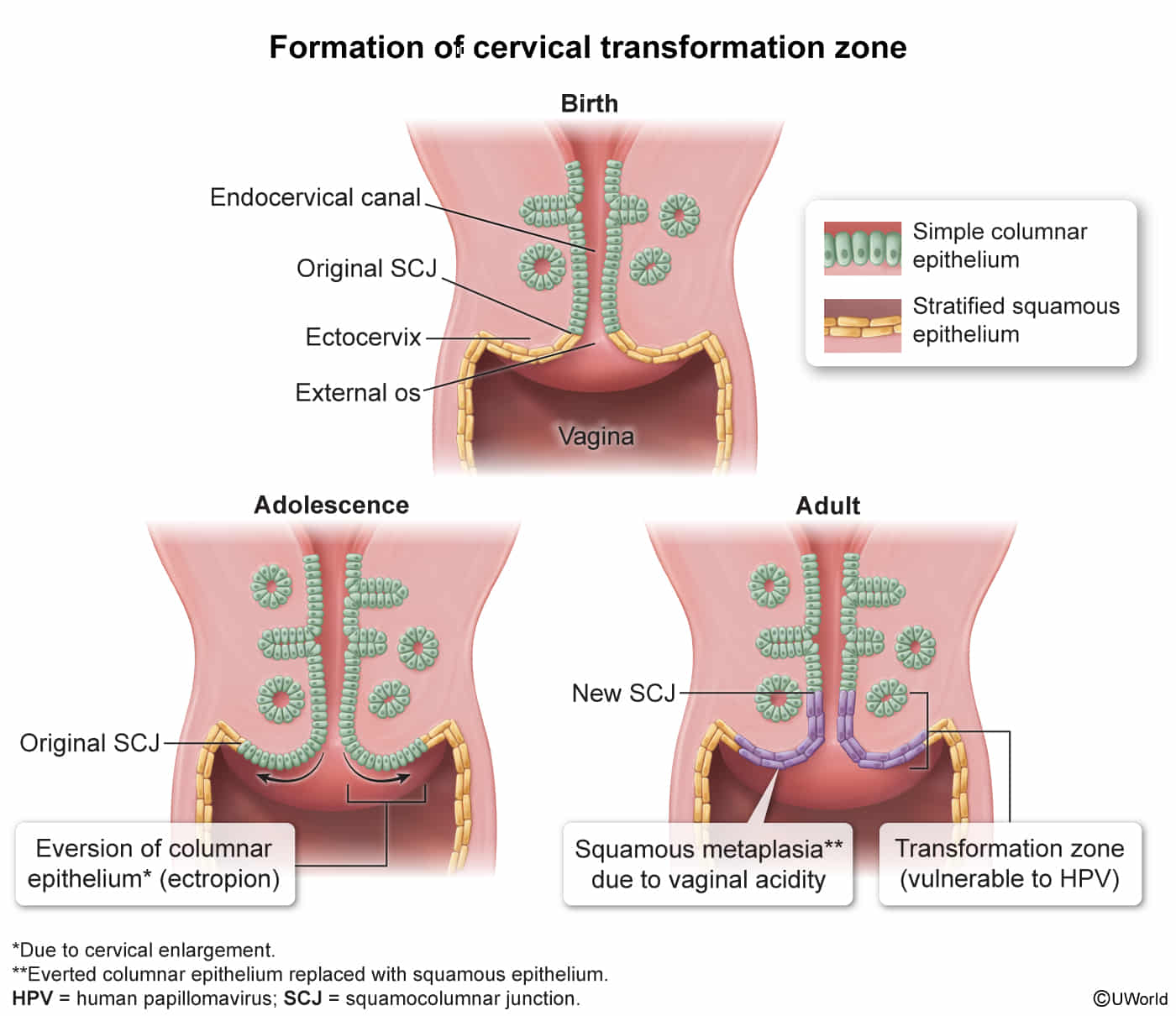
- A dynamic area on the ectocervix where columnar epithelium (ectropion) is gradually replaced by squamous epithelium through squamous metaplasia.
- This process is hormonally influenced and most active during puberty and first pregnancy.
- The TZ is the most common site for HPV infection and subsequent cervical dysplasia (CIN) and carcinoma.
- This is the critical area to sample during a Pap smear.
Clinical features
Diagnostics
| Feature | Cervical Cytology (Pap Smear Alone) | High-Risk HPV Co-Testing (Pap + hrHPV) |
|---|---|---|
| Primary Target | Phenotypic morphologic changes (dysplasia/SIL) | Presence of oncogenic viral DNA/RNA + cellular changes |
| Sensitivity for CIN 2/3+ | Moderate (50–70% for single test) | Very High (>90–95%) |
| Specificity for CIN 2/3+ | High (85–95%) | Lower in young patients; High in pts age ≥30 |
| Negative Predictive Value (NPV) | Moderate (requires shorter screening interval) | Near 100% (allows longer screening interval) |
| Recommended Screening Interval | Every 3 years | Every 5 years |
| Target Age Group | 21–65 years | 30–65 years (Not recommended <30 yrs) |
| Risk of Over-diagnosis / Overtreatment | Low | High in pts <30 yrs (detects transient infections) |
Differential diagnostics
| Feature | Cervical Cancer | Endometrial Cancer |
|---|---|---|
| Pathophysiology | HPV 16, 18 (E6 inhibits p53, E7 inhibits Rb). Arises at the transformation zone. | Unopposed estrogen leads to endometrial hyperplasia. |
| Classic Patient | Younger, multiple sexual partners, smoker, immunosuppressed (HIV). | Older, obese, nulliparous, diabetic. Postmenopausal. |
| Key Presentation | Postcoital bleeding. Often asymptomatic until advanced. | Postmenopausal bleeding. |
| Screening | Pap test +/- HPV co-testing. | None for asymptomatic women. |
| Diagnosis | Colposcopy with directed biopsy. | Endometrial biopsy. TVUS shows thickened stripe (>4mm). |
| Histology | Squamous Cell Carcinoma (most common). | Endometrioid adenocarcinoma (most common). |
| Major Risk Factors | - Multiple sexual partners - Immunosuppression - Smoking | - Obesity - Nulliparity - Unopposed estrogen therapy - Tamoxifen |
| Primary Treatment | Hysterectomy or chemoradiation (stage-dependent). | Total Hysterectomy + BSO. |
| Prevention | HPV vaccine. | Progestins (in combined OCPs/HRT), weight control. |
Pathology
- Invasive cervical carcinoma is characterized by invasion of the tumor beyond the basement membrane of the cervical epithelium.
- HSIL and invasive cervical carcinoma most commonly arise from metaplastic squamous cell epithelium in the cervical transformation zone
Squamous cell carcinoma (∼ 80% of cases)
- Subtypes include large cell keratinizing, large cell nonkeratinizing, and papillary squamous cell carcinoma.
- Irregular cell morphology
- Hyperchromatic cells with nonspherical nuclei, mitotic activity, and prominent nucleoli
- Loss of basal membrane
Tip
- Staging (Simplified FIGO)
- Stage I: Confined strictly to the cervix.
- Stage II: Invades beyond the uterus, but not to the pelvic wall or lower third of the vagina.
- Stage IIB is clinically important as it involves parametrial invasion.
- Stage III: Tumor extends to the pelvic wall, and/or involves the lower third of the vagina, and/or causes hydronephrosis.
- Stage IV: Extends beyond the true pelvis or involves mucosa of bladder/rectum (IVA) or has distant metastases (IVB).
Treatment
Pre-Invasive (CIN / HSIL)
- CIN 1:
- Observation (Preferred): Spontaneous resolution rate >80–90%.
- Age 21–24: Repeat Pap smear at 12 months.
- Age ≥25: Repeat HPV/co-test at 12 months. Treatment (LEEP/ablation) considered only if CIN 1 persists ≥2 years.
- CIN 2 & CIN 3:
- Excisional Treatment (First-line in non-pregnant adults ≥25 yo):
- LEEP (Loop Electrosurgical Excision Procedure): Preferred diagnostic & therapeutic method; removes transformation zone.
- CKC (Cold Knife Conization): Preferred if adenocarcinoma in situ (AIS), suspected microinvasion, unsatisfactory colposcopy, or positive ECC.
- Ablative Treatment (Cryotherapy, Laser ablation): Alternative only if squamocolumnar junction is fully visible and ECC is negative.
- Special Subpopulation (Ages 21–24 w/ CIN 2): Observation w/ colposcopy q6m for up to 24 months is acceptable to avoid future obstetrical complications, provided patient is compliant.
- Excisional Treatment (First-line in non-pregnant adults ≥25 yo):
- Post-Excision Surveillance (Post-LEEP / Post-CKC for CIN 2/3):
- First Follow-Up Test: hrHPV testing or co-testing at 6 months post-procedure. c
- If Negative (hrHPV negative / Cytology normal): Repeat hrHPV testing/co-testing annually for 3 consecutive years.
- If Positive (hrHPV (+) or Cytology ≥ASC-US): Diagnostic Colposcopy w/ ECC.
- Long-Term Surveillance (25-Year Rule):
- Following treatment for CIN 2, CIN 3, or AIS, patients require continued surveillance for at least 25 years (even if screening extends beyond age 65).
- Transition to routine screening (every 3 years for hrHPV/co-testing) only after passing initial post-treatment clearance.
- First Follow-Up Test: hrHPV testing or co-testing at 6 months post-procedure. c
- Management of Positive Margins or Positive Post-Excision ECC:
- Positive Margins w/ CIN 2/3: hrHPV testing at 6 months is preferred; repeat excision is reserved for persistent disease or if colposcopy/ECC is inadequate.
- Positive Margins w/ Adenocarcinoma in Situ (AIS): Repeat excisional procedure (CKC) to ensure negative margins (high risk of skip lesions).
Invasive Cancer
- Stage IA1 (Microinvasive, <3 mm depth):
- Cone biopsy w/ negative margins (if fertility preservation desired).
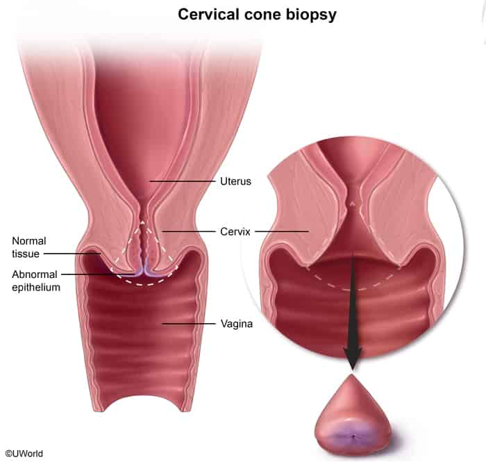
- Simple Hysterectomy (if fertility not desired).
- Cone biopsy w/ negative margins (if fertility preservation desired).
- Stage IA2 - IB2 / IIA1 (Early / Operable, ≤4 cm, no parametrial invasion):
- Radical Hysterectomy + Pelvic Lymphadenectomy OR
- Definitive Chemoradiation (equivalent survival outcomes).
- Stage IIB - IVA (Locally Advanced, parametrial invasion, lower 1/3 vagina, or bladder/rectum):
- Definitive Concurrent Chemoradiation (Cisplatin-based chemotherapy + EBRT + Brachytherapy).
- Surgical resection is contraindicated.
- Stage IVB (Distant Metastasis):
- Palliative chemotherapy (e.g., Cisplatin + Paclitaxel + Bevacizumab) + local palliative EBRT.
Complications
- Complications of Excisional Procedures (LEEP/CKC):
- Cervical insufficiency (incompetence) → increased risk of preterm delivery and 2nd-trimester pregnancy loss. c
- Cervical stenosis → dysmenorrhea, hematometra, and subfertility.
- Intraoperative/postoperative hemorrhage and localized pelvic infection.
Screening
- Standard Screening Protocol:
- Age < 21: No screening (regardless of sexual history).
- Age 21–29: Cytology (Pap) alone q3y (no routine HPV testing due to high prevalence of transient HPV infections).
- Age 30–65: Co-testing (Pap + hrHPV) q5y (preferred), hrHPV alone q5y, or Pap alone q3y. c
- Age > 65: Discontinue if adequate prior screening (3 consecutive (-) Paps or 2 consecutive (-) co-tests within past 10 yr) AND no hx of CIN 2+ in past 25 yr.
- Because progression from a new hrHPV infection to invasive cervical carcinoma takes 10–20+ years.
- Special Populations:
- Total hysterectomy (benign): Discontinue screening.
- Supracervical hysterectomy: Continue routine screening.
- Hx of CIN 2/3 or invasive CA: Continue screening for 25 years post-treatment.
- Immunocompromised (HIV, transplant): Start at sexual debut/diagnosis annual Pap , then q3y.
- HPV vaccinated: Follow the exact same routine screening guidelines.
- Abnormal Result Management:
- ASC-US:
- Age 21–24: Repeat Pap in 12 months.
- Age : Reflex HPV if (+), Colposcopy; if (-), repeat co-test in 3 yr.
- LSIL:
- Age 21–24: Repeat Pap in 12 months.
- Age : Colposcopy.
- ASC-H: Colposcopy for all age groups.
- HSIL: Colposcopy (or immediate LEEP/conization if age ).
- AGC: Colposcopy + ECC + EMB (EMB if age or high risk for endometrial CA).
- Benign Endometrial Cells on Pap:
- Premenopausal: Normal finding; no workup needed.
- Postmenopausal: Perform Endometrial Biopsy (EMB) to rule out endometrial hyperplasia/carcinoma. c
- Because the atrophic endometrium does not undergo cyclic turnover, it should not shed cells. Exfoliation of endometrial cells indicates abnormal tissue fragility or proliferation
- ASC-US:
Cervical cancer - abnormal Pap and HPV management.excalidraw
⚠ Switch to EXCALIDRAW VIEW in the MORE OPTIONS menu of this document. ⚠ You can decompress Drawing data with the command palette: ‘Decompress current Excalidraw file’. For more info check in plugin settings under ‘Saving’
Excalidraw Data
Text Elements
Abnormal Pap Smear Cytology +/- Abnormal hrHPV Co-testing
LSIL
ASC-US
ASC-H
HSIL
SCC (clear evidence)
Atypical glandular cells
Endometrial cells
Reflex hrHPV testing
Repeat testing in 1 year (age 21-24) or co-testing in 3 years (age >24)
Colposcopy +/- biopsy
Mildly atypical cells in the lower 1/3 of cervix
Moderately atypical cells in the lower 2/3 of cervix
Severely atypical cells in more than the lower 2/3 of cervix
Mild dysplasia (CIN I)
Moderate dysplasia (CIN II)
Severe dysplasia (CIN III)
Repeat testing in 1 year
LEEP or conization
LEEP or conization
Squamous cell carcinoma
Surgical resection and/or chemotherapy (stage-dependent)
Endometrial sampling (if age >=35 or high risk)
CIN II or less
Non-diagnostic or evidence of AIS
Repeat Pap smear with hrHPV co-testing in 12 months
Conization
Adenocarcinoma in situ (AIS)
Adenocarcinoma
Surgical resection
Surgical resection
Endometrial sampling and endocervical curettage
Endometrial pathology
No endometrial pathology
See Abnormal Uterine Bleeding
Proceed based on diagnostic findings
Colposcopy with endocervical curettage
Colposcopy
Element Links
aub-box: Abnormal uterine bleeding
Link to original
- Diagnostic & Excisional Procedures
- Colposcopy & Cervical Biopsy:
- Application of 3–5% acetic acid (highlights acetowhite changes) and Lugol iodine (normal tissue stains dark brown; abnormal fails to stain).
- Satisfactory Colposcopy: Entire transformation zone (squamocolumnar junction) is completely visualized.
- Unsatisfactory Colposcopy: Squamocolumnar junction not fully seen → perform Endocervical Curettage (ECC). c
- Endocervical Curettage (ECC):
- Evaluates the endocervical canal when lesions extend into the os or transformation zone is obscured.
- Colposcopy & Cervical Biopsy:
- Pregnancy Management:
- Colposcopy and cervical biopsy are safe if invasive CA is suspected.
- Contraindicated in pregnancy: ECC, EMB, and routine excisional procedures (LEEP/CKC).
- High-Yield Clinical Pearls:
- Grossly visible cervical mass / postcoital bleeding: Do not perform Pap or colposcopy Immediate punch biopsy.
- Conization complications: Cervical insufficiency (leading to preterm delivery/loss) and cervical stenosis.
- HPV 16 & 18: Main oncogenic strains (HPV 16 squamous cell CA; HPV 18 adenocarcinoma).