Atherosclerosis

Definitions

  • Arteriosclerosis: an umbrella term to describe arterial wall thickening (hardening) and elasticity loss with variable pathogenesis
  • Atherosclerosis
    • Most common type of arteriosclerosis
    • Multifactorial inflammatory disease of the intima, manifesting at points of hemodynamic shear stress
    • Characterized by a build-up of cholesterol plaques in the tunica intima
    • Affects elastic arteries and large/medium-sized muscular arteries
  • Monckeberg arteriosclerosis
    • A form of arteriosclerosis characterized by dystrophic calcification of the tunica media and internal elastic lamina
    • There is no blood flow obstruction.
      • The intimal layer of the artery is not involved.
    • Mainly affects medium-sized arteries
    • Causes: diabetes mellitus and/or progressive kidney disease
    • X-ray: pipestem appearance
  • Arteriolosclerosis: the thickening and loss of elasticity of the small arteries and arterioles
    • Hyaline arteriolosclerosis
      • Deposition of proteins below the endothelium due to plasma protein leakage
      • Causes: chronic essential hypertension, diabetes mellitus, and normal aging
      • H&E: pink amorphous deposits (hyaline) within the arteriolar walls
    • Hyperplastic arteriolosclerosis
      • Proliferation of subendothelial smooth muscle cells in response to very high blood pressure
      • Cause: malignant hypertension
      • H&E: “onion-skin” appearance of the arteriole

Pathogenesis of atherosclerosis

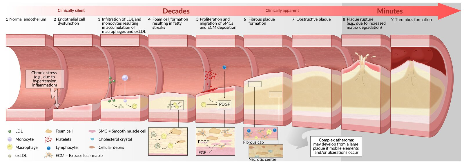
  1. Chronic stress on the endothelium (e.g., due to arterial hypertension and turbulence)
  2. Endothelial cell dysfunction, which leads to:
    • Invasion of inflammatory cells (mainly monocytes and lymphocytes) through the disrupted endothelial barrier
    • Adhesion of platelets to the damaged vessel wall → platelet release of inflammatory mediators (e.g., cytokines) and platelet-derived growth factor (PDGF) t
    • PDGF stimulates the migration and proliferation of smooth muscle cells (SMCs) in the tunica intima and mediates the differentiation of fibroblasts into myofibroblasts
  3. Inflammation of the vessel wall
  4. Macrophages and SMCs ingest cholesterol from oxidized LDL and transform into foam cells (macrophages filled with lipid droplets).
  5. Foam cells accumulate to form fatty streaks (early atherosclerotic lesions).
  6. Lipid-laden macrophages and SMCs produce extracellular matrix (e.g., collagen) deposition → development of a fibrous plaque (atheroma)
    • Macrophages, smooth muscle cells, lymphocytes, and extracellular matrix form a fibrous cap, which covers a necrotic center, consisting of foam cells, free cholesterol crystals, and cellular debris.
  7. Inflammatory cells in the atheroma (e.g., macrophages) secrete matrix metalloproteinases → weakening of the fibrous cap of the plaque due to the breakdown of extracellular matrix → minor stress ruptures the fibrous cap
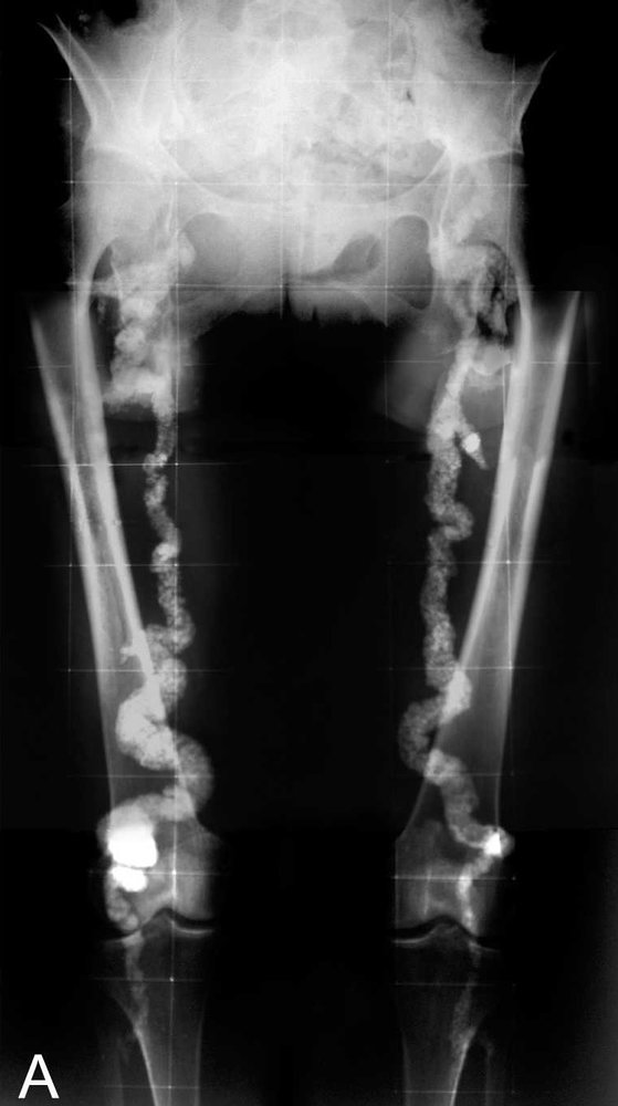
  8. Calcification of the intima (the amount and pattern of calcification affect the risk of complications)
  9. Plaque rupture → exposure of thrombogenic material (e.g., collagen) → thrombus formation with vascular occlusion or spreading of thrombogenic material

Diagnostics

  • Initial/Screening (Primary Care Focus):
    • 10-year ASCVD Risk Estimator (Pooled Cohort Equations) for adults aged 40-75.
    • Fasting lipid panel, HbA1c/Fasting glucose, BP measurement.
  • Bed-Specific Testing:
    • CAD: Resting ECG, Stress testing (Exercise ECG, Echo, or Nuclear), Coronary CT Angiography (CCTA).
    • PAD: Ankle-Brachial Index (ABI) ≤ 0.90 is diagnostic.
    • CVA: Carotid duplex ultrasound, CTA/MRA of head/neck.
  • Confirmatory/Gold Standard: Invasive Angiography (Coronary, Peripheral, or Cerebral) — typically reserved for planned intervention/revascularization.

Management

  • Primary Prevention (No established ASCVD):
    • Lifestyle: Smoking cessation, Mediterranean/DASH diet, weight loss, exercise.
    • BP Control: Target <130/80 mmHg per ACC/AHA guidelines.
    • Glycemic Control: Target HbA1c <7% for most DM pts (SGLT2i/GLP-1 RA preferred for high CV risk).
    • Statin Indications: c
      1. LDL ≥ 190 mg/dL: High-intensity statin.
      2. DM (Age 40-75): Moderate-intensity statin (High-intensity if multiple risk factors).
      3. 10-year ASCVD risk ≥ 7.5% - 20% (Age 40-75): Moderate-intensity statin.
      4. 10-year ASCVD risk ≥ 20%: High-intensity statin.
    • Aspirin: Routine primary prevention no longer recommended for adults >60 due to bleeding risk. Consider only in select high-risk adults 40-59 w/ low bleeding risk.
  • Secondary Prevention (Known ASCVD):
    • Antiplatelet: Aspirin 81 mg daily. Dual Antiplatelet Therapy (DAPT) w/ P2Y12 inhibitor (e.g., Clopidogrel, Ticagrelor) for 6-12 months post-ACS or PCI.
    • High-Intensity Statin: Atorvastatin 40-80 mg or Rosuvastatin 20-40 mg daily (Target LDL < 70 mg/dL). Add Ezetimibe or PCSK9i if target not met.
    • Beta-Blockers: Indicated for Hx of MI, stable angina, or HFrEF.
    • ACEi/ARB: Indicated for HTN, DM, CKD, or HFrEF (EF ≤ 40%).
  • Revascularization: PCI/CABG for CAD; Endarterectomy/Stenting for carotids; PTA/Bypass for PAD.