| Feature | Stress | Urge (OAB) | Overflow |
|---|---|---|---|
| Mechanism | Urethral Hypermobility or Sphincter deficiency | Detrusor Overactivity (uninhibited spasms) | Impaired Contractility (atonic) or Obstruction |
| Symptoms | Leak w/ ↑ abd pressure (cough, sneeze, lift) | Sudden, overwhelming need to void | Constant dribbling c, incomplete emptying |
| Key Risk | Multiparous women, Obesity | Elderly, MS, Idiopathic | BPH, Diabetes (neurogenic), Spinal injury |
| Diagnosis | + Bladder Stress Test (visual leak) | Clinical diagnosis | High PVR (>200mL) |
| Tx: 1st Line | Kegels (Pelvic floor training) | Bladder training | Catheterization (immediate relief) |
| Tx: Meds | (None effective) | Oxybutynin (Antimuscarinic), Mirabegron (β3-agonist) | Tamsulosin (α-blocker), Bethanechol (cholinergic) |
| Tx: Surg | Mid-urethral Sling | Botox (refractory) | TURP (if BPH) |
Mixed Incontinence: Features of both stress and urge incontinence. Very common, especially in older women. Treatment is directed at the more predominant symptom first.
Functional Incontinence: Patient has normal bladder control but cannot reach the toilet in time due to physical or cognitive impairment (e.g., dementia, severe arthritis). Management involves treating the underlying condition and using scheduled/prompted voiding.
Epidemiology
- Stress incontinence and mixed incontinence are the most common types of incontinence in female patients.
- Urge incontinence is the most common type in male patients.
Etiology
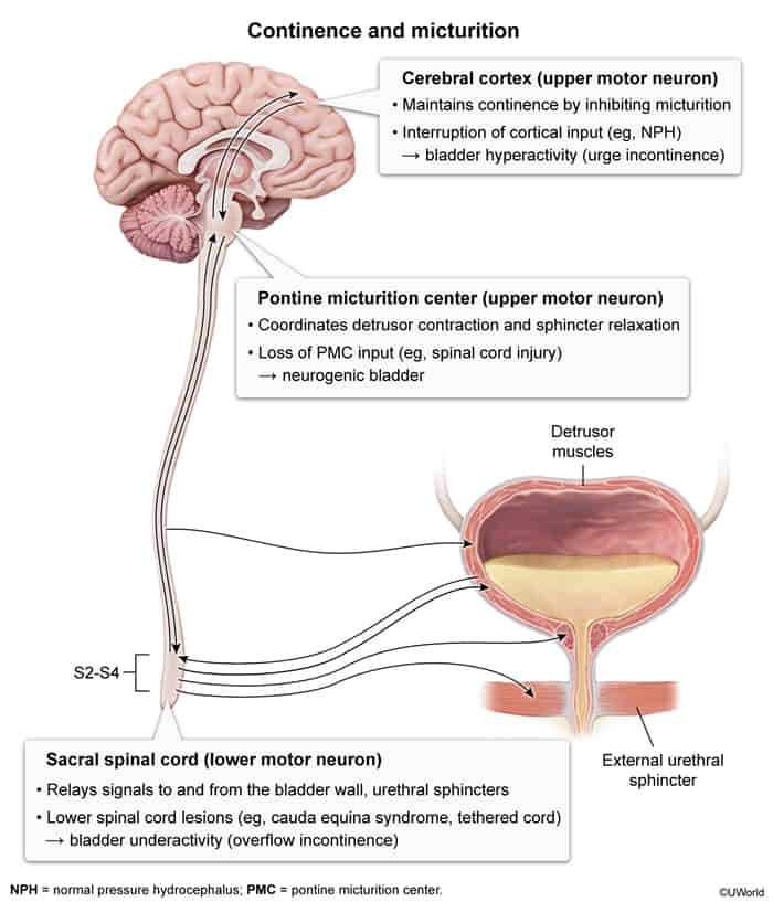
- Neurological causes
- Multiple sclerosis
- Spinal injury
- Normal pressure hydrocephalus
- Genitourinary causes
- Trauma to the pelvic floor
- Intrinsic sphincter deficiency
- Urethral hypermobility in women
- Impaired detrusor contractility
- Bladder outlet obstruction
- Pelvic floor weakness
- Transient causes of urinary incontinence
- Drugs (e.g., diuretics)
- Urinary tract infections
- Postmenopausal atrophic urethritis
- Psychiatric causes (especially depression, delirium/confused state)
- Excessive urinary output (in conditions like hyperglycemia, hypercalcemia, CHF)
- Stool impaction
- General risk factors
- Recurrent urinary tract infections
urethritis/vaginitis, Pharmaceutical, Psychiatric causes (especially depression), Excessive urinary output (hyperglycemia, hypercalcemia, CHF), Restricted mobility, Stool impaction.
To remember the reversible causes of acute urinary incontinence, think DIAPPERS: Delirium/confusion, Infection, Atrophic
Classifications
Overflow incontinence
Mechanism
- Impaired (weak) detrusor contractility
- Bladder outlet obstruction (e.g., BPH)
- Both mechanisms can lead to incomplete bladder emptying → bladder overfilling → chronically distended bladder with ↑ bladder pressure → dribbling of urine (leak) when intravesical pressure > outlet resistance
Clinical features
- Frequent, involuntary intermittent/continuous dribbling of urine in the absence of an urge to urinate
- Occurs only when the bladder is full
- Postvoid residual urine volume (seen on ultrasound or with catherization)
- Assess for urinary retention via bladder scan (≥200 mL) or straight catheterization yielding ≥200 mL urine output.
Treatment
- Urinary catheter: for scheduled bladder emptying
- Intermittent -> prevention of catheter-associated UTIs and is favored by most patients; indwelling catheters may be considered if necessary.
Stress incontinence
Epidemiology
Prevalence increases with age.
Etiology
- Pathophysiology:
- Urethral hypermobility: Weak pelvic floor musculature/fascial support fails to support the bladder neck and urethra under increased intra-abdominal pressure. c
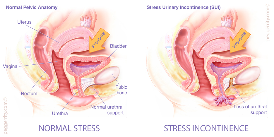
- Intrinsic sphincteric deficiency (ISD): Loss of intrinsic urethral mucosal coaptation/sphincter function (e.g., trauma, multiple pelvic surgeries, pelvic radiation).
- Risk Factors:
- Multiparity / Vaginal delivery (major risk factor for pelvic floor trauma/nerve stretch injury).
- Obesity (chronically ↑ intra-abdominal pressure).
- Advanced age & Postmenopausal state (hypoestrogenism leads to urogenital atrophy).
- Prior pelvic surgery (e.g., hysterectomy).
- Chronic conditions causing increased abdominal pressure (COPD/chronic cough, chronic constipation).
Clinical features
- History:
- Involuntary, instantaneous loss of urine with exertion or increased intra-abdominal pressure (e.g., coughing, sneezing, laughing, lifting, jumping).
- No preceding urge sensation or sensation of bladder fullness prior to leakage.
- Physical Exam:
- Positive Cough Stress Test: Direct visualization of transurethral urine loss simultaneously with coughing/Valsalva in the lithotomy or standing position.
- Pelvic Exam: May demonstrate concomitant pelvic organ prolapse (POP), such as cystocele, rectocele, or uterine prolapse; vaginal mucosal atrophy. c
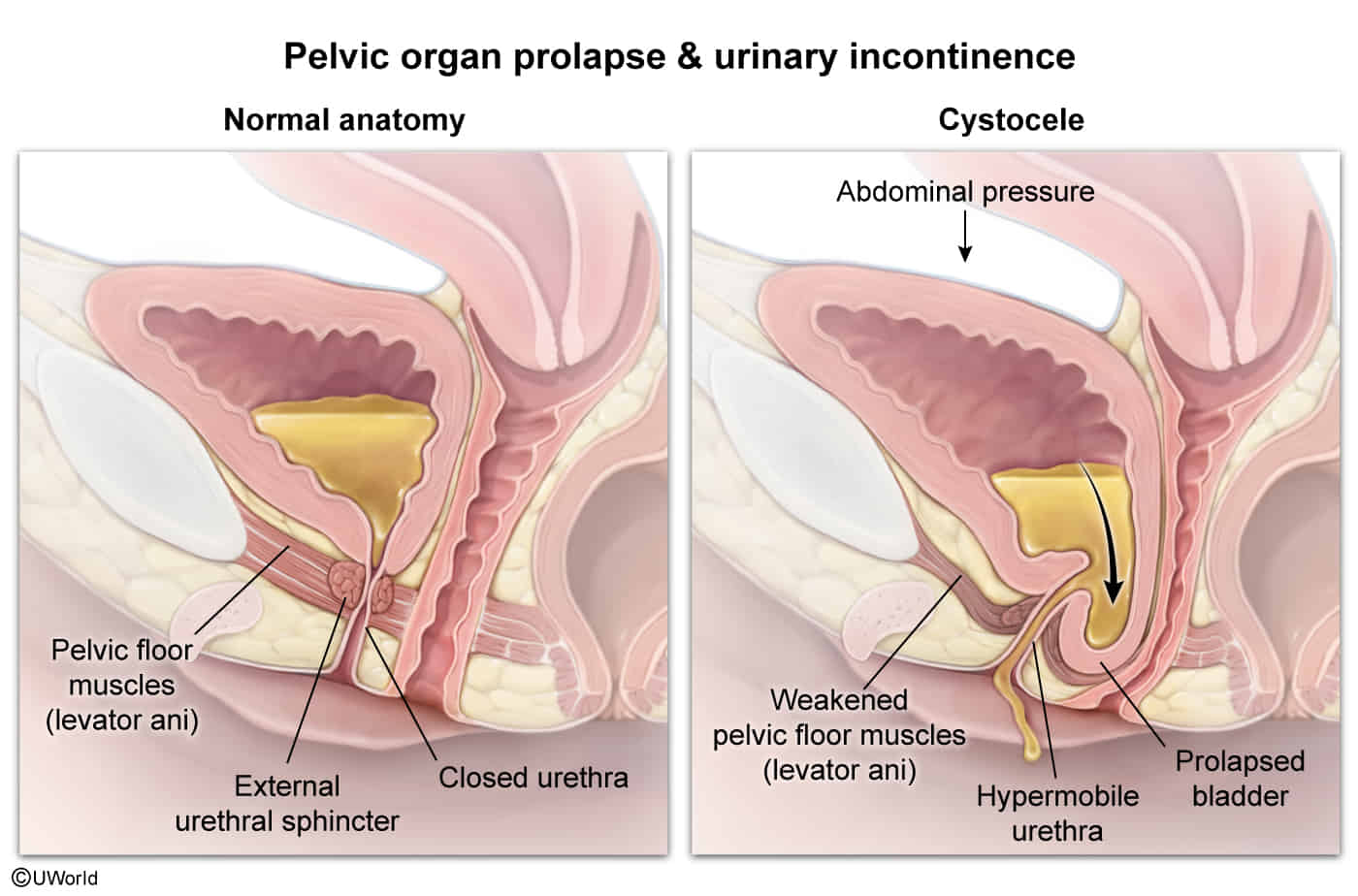
- Q-tip test: Swab deflection > 30 degrees from horizontal with Valsalva indicates urethral hypermobility.
Diagnostics
Treatment
- First-Line (Conservative / Non-invasive):
- Pelvic Floor Muscle Training (PFMT / Kegel exercises): Strengthens levator ani muscles (pubococcygeus); first-line therapy for all patients.
- Lifestyle Modifications: Weight loss (highly effective), fluid management, smoking cessation, optimization of chronic cough and constipation.
- Second-Line (Non-surgical Mechanical Support):
- Pessary (e.g., incontinence dish or ring pessary with knob): Elevates and supports the bladder neck; ideal for patients who decline surgery, are pregnant, or are poor surgical candidates.
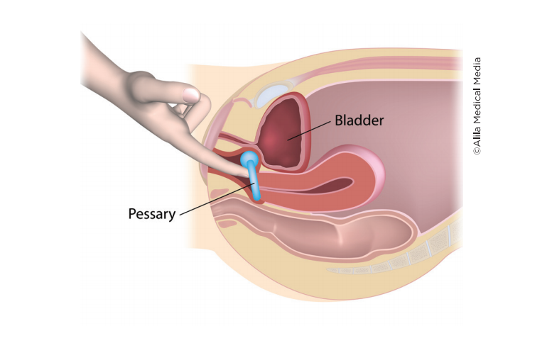
- Vaginal inserts / Urethral plugs: Temporary mechanical support during high-impact activities.
- Definitive / Surgical Management (Indicated for severe or refractory symptoms):
- Midurethral Sling (Gold Standard): Placement of a synthetic mesh sling (e.g., tension-free vaginal tape [TVT] or transobturator tape [TOT]) under the mid-urethra to restore backboard support.
- Bladder Neck Suspension (Burch Colposuspension): Open or laparoscopic suspension of the periurethral tissue to Cooper’s ligament.
- Periurethral / Transurethral Bulking Agents: Injection of cross-linked collagen or synthetic gel into the submucosa around the bladder neck; preferred for intrinsic sphincteric deficiency in high-risk surgical patients.
- Note on Pharmacotherapy: Systemic medications (e.g., alpha-agonists, duloxetine) have low efficacy and significant adverse effects; not standard first-line therapy in US guidelines.
Link to original
Stress incontinence
Epidemiology
Prevalence increases with age.
Etiology
- Pathophysiology:
- Urethral hypermobility: Weak pelvic floor musculature/fascial support fails to support the bladder neck and urethra under increased intra-abdominal pressure. c
- Intrinsic sphincteric deficiency (ISD): Loss of intrinsic urethral mucosal coaptation/sphincter function (e.g., trauma, multiple pelvic surgeries, pelvic radiation).
- Urethral hypermobility: Weak pelvic floor musculature/fascial support fails to support the bladder neck and urethra under increased intra-abdominal pressure. c
- Risk Factors:
- Multiparity / Vaginal delivery (major risk factor for pelvic floor trauma/nerve stretch injury).
- Obesity (chronically ↑ intra-abdominal pressure).
- Advanced age & Postmenopausal state (hypoestrogenism leads to urogenital atrophy).
- Prior pelvic surgery (e.g., hysterectomy).
- Chronic conditions causing increased abdominal pressure (COPD/chronic cough, chronic constipation).
Clinical features
- History:
- Involuntary, instantaneous loss of urine with exertion or increased intra-abdominal pressure (e.g., coughing, sneezing, laughing, lifting, jumping).
- No preceding urge sensation or sensation of bladder fullness prior to leakage.
- Physical Exam:
- Positive Cough Stress Test: Direct visualization of transurethral urine loss simultaneously with coughing/Valsalva in the lithotomy or standing position.
- Pelvic Exam: May demonstrate concomitant pelvic organ prolapse (POP), such as cystocele, rectocele, or uterine prolapse; vaginal mucosal atrophy. c
- Q-tip test: Swab deflection > 30 degrees from horizontal with Valsalva indicates urethral hypermobility.
Diagnostics
Treatment
- First-Line (Conservative / Non-invasive):
- Pelvic Floor Muscle Training (PFMT / Kegel exercises): Strengthens levator ani muscles (pubococcygeus); first-line therapy for all patients.
- Lifestyle Modifications: Weight loss (highly effective), fluid management, smoking cessation, optimization of chronic cough and constipation.
- Second-Line (Non-surgical Mechanical Support):
- Pessary (e.g., incontinence dish or ring pessary with knob): Elevates and supports the bladder neck; ideal for patients who decline surgery, are pregnant, or are poor surgical candidates.
- Vaginal inserts / Urethral plugs: Temporary mechanical support during high-impact activities.
- Pessary (e.g., incontinence dish or ring pessary with knob): Elevates and supports the bladder neck; ideal for patients who decline surgery, are pregnant, or are poor surgical candidates.
- Definitive / Surgical Management (Indicated for severe or refractory symptoms):
- Midurethral Sling (Gold Standard): Placement of a synthetic mesh sling (e.g., tension-free vaginal tape [TVT] or transobturator tape [TOT]) under the mid-urethra to restore backboard support.
- Bladder Neck Suspension (Burch Colposuspension): Open or laparoscopic suspension of the periurethral tissue to Cooper’s ligament.
- Periurethral / Transurethral Bulking Agents: Injection of cross-linked collagen or synthetic gel into the submucosa around the bladder neck; preferred for intrinsic sphincteric deficiency in high-risk surgical patients.
- Note on Pharmacotherapy: Systemic medications (e.g., alpha-agonists, duloxetine) have low efficacy and significant adverse effects; not standard first-line therapy in US guidelines.
Urge incontinence
Epidemiology
Prevalence increases with age
Etiology
The condition is caused by sensory or motor dysfunction.
- Idiopathic (most common)
- Neurological conditions
Pathophysiology
Inflammatory conditions (e.g., UTI) or neurogenic disorders → sphincter dysfunction, detrusor overactivity, or overactive bladder → autonomous contractions of the detrusor muscle and premature initiation of a normal micturition reflex
Clinical features
- Urinary urgency: sudden urge to urinate
- Loss of urine without exertion, with urinary tenesmus → frequent episodes, with variable volumes of urine voided each time
Diagnostics
Treatment
Mnemonic
Oxybutynin treats Overactive bladder.
Link to original
Urge incontinence
Epidemiology
Prevalence increases with age
Etiology
The condition is caused by sensory or motor dysfunction.
- Idiopathic (most common)
- Neurological conditions
Pathophysiology
Inflammatory conditions (e.g., UTI) or neurogenic disorders → sphincter dysfunction, detrusor overactivity, or overactive bladder → autonomous contractions of the detrusor muscle and premature initiation of a normal micturition reflex
Clinical features
- Urinary urgency: sudden urge to urinate
- Loss of urine without exertion, with urinary tenesmus → frequent episodes, with variable volumes of urine voided each time
Diagnostics
Treatment
Mnemonic
Oxybutynin treats Overactive bladder.
Link to original
Neurogenic bladder dysfunction
Mechanism
detrusor sphincter dyssynergia
- Simultaneous contractions of the detrusor muscle and involuntary activation of the urethral sphincter → blockage of bladder outlet → small amounts of urine are pressed through the contracted sphincter muscle → high intravesical pressure along with inappropriate contraction of the urethral sphincter
- Commonly seen in multiple sclerosis and spinal cord injury
Clinical features
- Voiding and/or storage dysfunction, intermittent voiding, urinary retention
- Irregular, small volume incontinence without an associated urge to void (sometimes referred to as reflex incontinence)
Total incontinence
Complete loss of sphincter function (due to previous surgery, nerve damage, metastasis) → Urinary leakage occurs at all times
Clinical features
Diagnostics
Treatment
Autonomic & Somatic Innervation
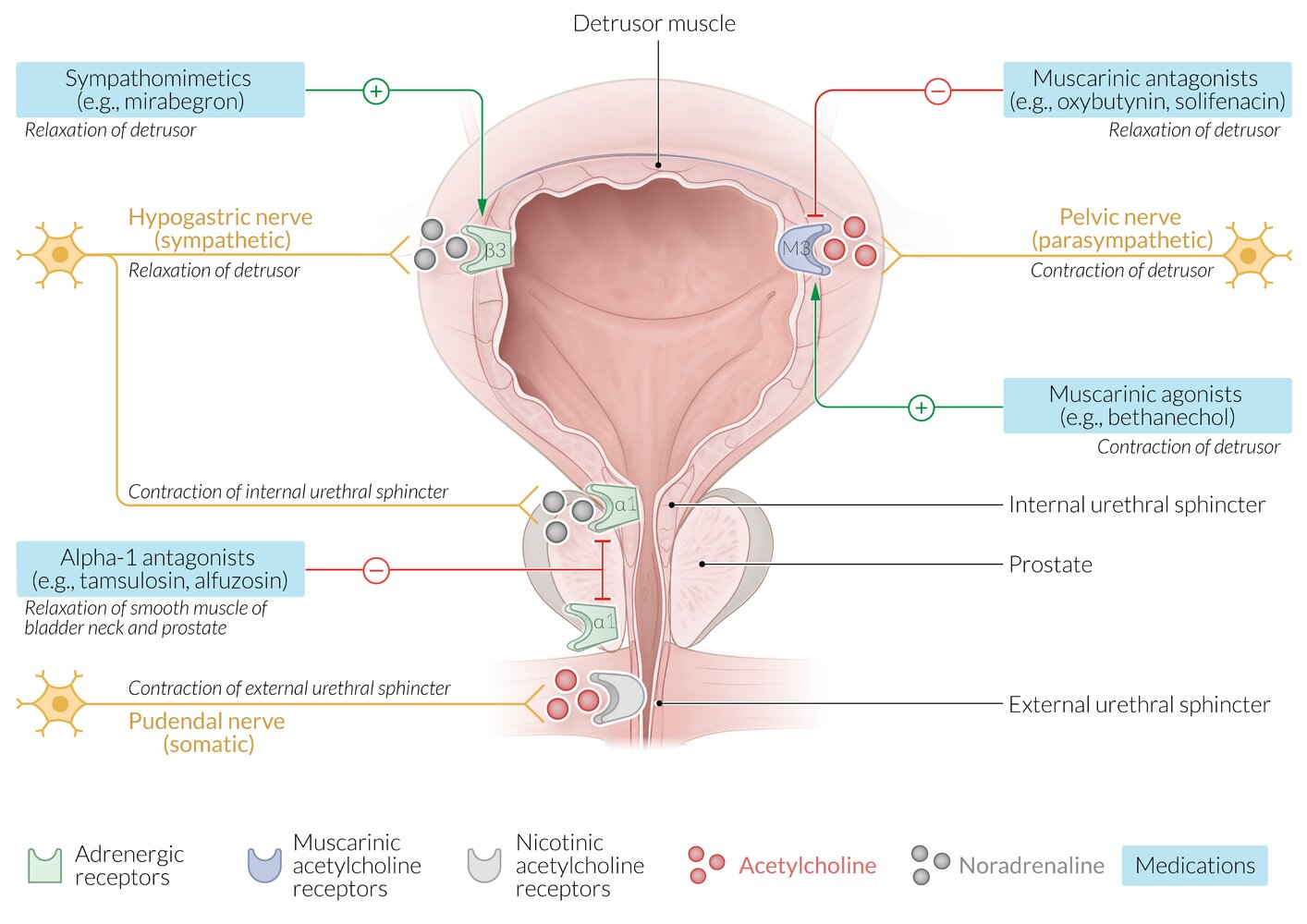
- Sympathetic (Storage/“Sympathetic Stores”): Hypogastric nerve (T11-L2).
- Relaxes detrusor muscle (via β3 receptors) → allows filling.
- Contracts internal urethral sphincter (via α1 receptors) → prevents leakage.
- Parasympathetic (Peeing/Voiding): Pelvic splanchnic nerves (S2-S4).
- Contracts detrusor muscle (via M3 receptors) → initiates emptying.
- Relaxes internal urethral sphincter (via M3 receptors).
- Somatic (Voluntary Control): Pudendal nerve (S2-S4).
- Contracts external urethral sphincter (via Nicotinic receptors) → allows voluntary holding of urine.
Overview of pharmacotherapy
Warning
The use of muscarinic agonists may lead to urinary urgency, while the use of sympathomimetics or muscarinic antagonists may lead to urinary retention, especially if there is an untreated outlet obstruction.
Tip
No pharmacological therapies are FDA-approved for stress incontinence; treatment is primarily conservative with surgery.