Key points
- Multiple sclerosis (MS) is a chronic degenerative disease of the CNS characterized by demyelination and axonal degeneration in the brain and spinal cord, which are caused by an immune-mediated inflammatory process.
- Impaired vision (due to retrobulbar neuritis) is usually the first manifestation of MS; other neurological deficits appear as the disease progresses. The most common clinical course is characterized by exacerbations followed by periods of complete or incomplete remission.
Epidemiology
- Sex: ♀ > ♂ (3:1)
- Age of onset: 20–40 years of age
- Ethnicity: ↑ prevalence among the white and black population
- Prevalence: 50-300 per 100 000 people (greater among people who live further from the equator)
Etiology
The etiology of multiple sclerosis is unclear; it is believed to develop in genetically predisposed people who have been exposed to certain environmental factors.
- Environmental risk factors
Pathophysiology
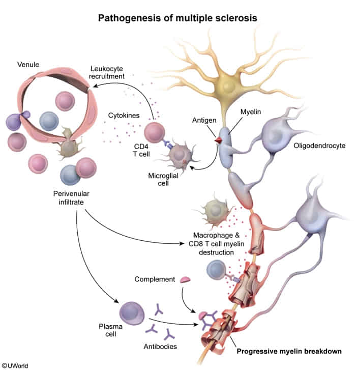
- Autoreactive T cells cross BBB → attack myelin
- Plaques (sclerotic lesions) form in periventricular white matter, optic nerves, brainstem, spinal cord
- Demyelination → ↓ conduction velocity → neurologic deficits
- Acute: inflammation, demyelination
- Chronic: gliosis, axonal loss (irreversible)
Tip
Only CNS demyelination, no PNS demyelination (unlike GBS) c
Clinical features
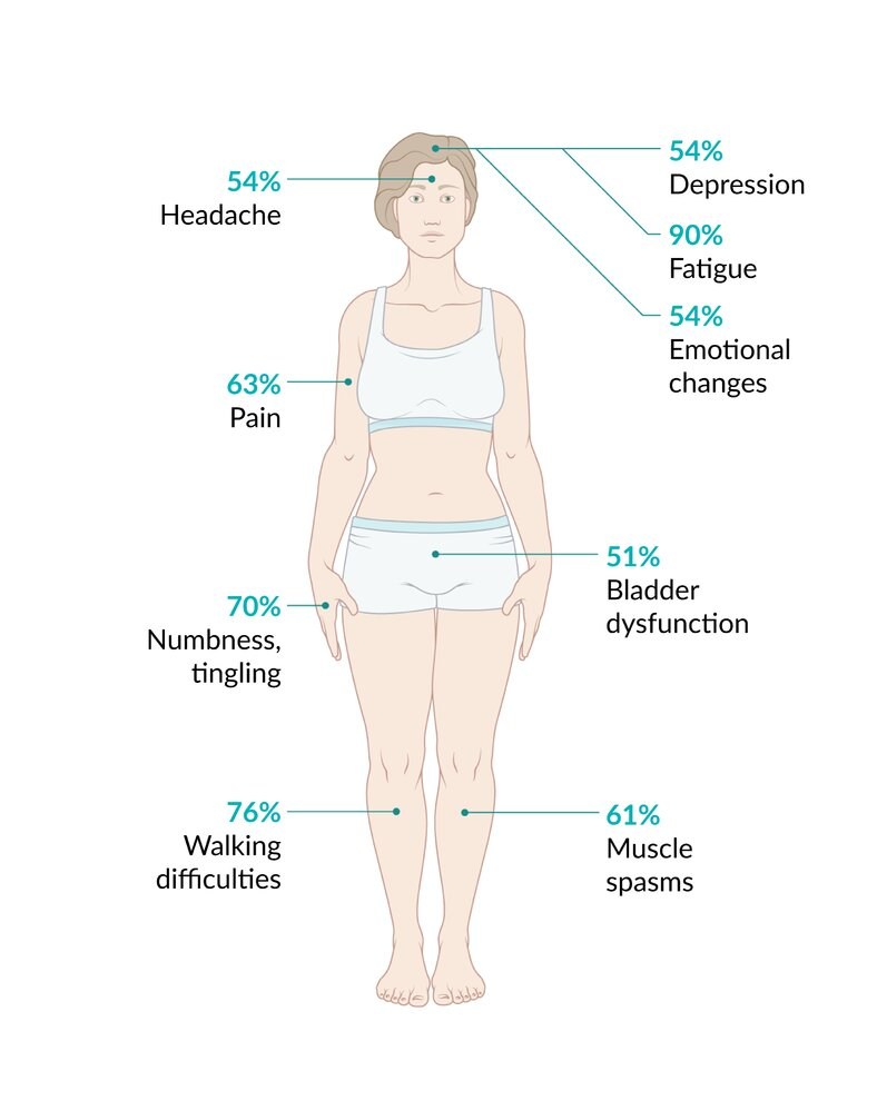
- Core Feature: Multiple neurological deficits disseminated in space and time (CNS demyelinating lesions separated by anatomic location and chronological occurrence). c
- Sensory Disturbances:
- Paresthesias, numbness, decreased vibration/proprioception.
- Lhermitte sign: Electric shock-like sensation radiating down the spine and into the limbs upon neck flexion. c
- Motor Symptoms:
- Upper motor neuron (UMN) signs: Spasticity, hyperreflexia, (+) Babinski sign, muscle weakness.
- Pronator drift: Sensitive physical exam marker for UMN / corticospinal tract lesion (pt extends arms with palms up and eyes closed affected arm pronates and drifts downward due to weakness in supinator muscles relative to pronators). c
- Upper motor neuron lesions cause more weakness in the supinator muscles compared to the pronator muscles of the upper limb.
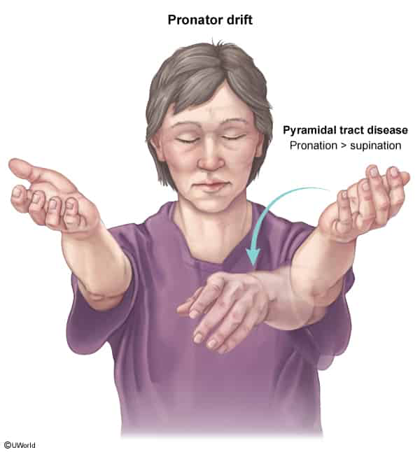
- Upper motor neuron lesions cause more weakness in the supinator muscles compared to the pronator muscles of the upper limb.
- Ocular & Visual Symptoms:
- Optic Neuritis: Acute, unilateral, painful eye movement associated with central vision loss, central scotoma, impaired color vision (dyschromatopsia), and afferent pupillary defect (Marcus Gunn pupil).
- Internuclear Ophthalmoplegia (INO): Damage to the Medial Longitudinal Fasciculus (MLF). Impaired adduction of the ipsilateral eye with horizontal nystagmus of the contralateral abducting eye during lateral gaze; convergence is preserved. Bilateral INO is highly suggestive of MS.
- Cerebellar & Brainstem Symptoms:
- Charcot neurologic triad: Scanning speech (dysarthria), Intention tremor, Nystagmus.
- Ataxia, vertigo, scanning dysarthria.
- Autonomic & Other Features:
- Uhthoff phenomenon: Transient worsening of neurological symptoms with increased body temperature (e.g., hot shower, exercise, fever).
- Impulse conduction is dependent on temperature. An increase in body temperature presumably worsens impulse conduction in demyelinated nerves.
- Neurogenic bladder: Spastic/hyperreflexic bladder causing urge incontinence (detrusor hyperreflexia) or flaccid bladder causing overflow retention. c
- Severe chronic fatigue, depression, cognitive dysfunction.
- Depression has been found in up to two-thirds of patients with MS and is the most common psychiatric complication. c
- Uhthoff phenomenon: Transient worsening of neurological symptoms with increased body temperature (e.g., hot shower, exercise, fever).
- Disease Courses:
- Relapsing-Remitting (RRMS): Most common (~85%); acute attacks with full or partial recovery, stable between attacks.
- Secondary Progressive (SPMS): Initial RRMS followed by gradual progression with or without relapses.
- Primary Progressive (PPMS): Continuous progression of disability from onset (~15%).
Tip
MS is a chronic condition that typically manifests in a relapsing-remitting form characterized by episodic CNS dysfunction (exacerbations) with at least partial recovery between episodes.
Diagnostics
- Initial & Confirmatory / Gold Standard: Brain and Spinal Cord MRI with and without IV Gadolinium. c
- T2/FLAIR: Hyperintense demyelinating plaques in periventricular regions oriented perpendicular to the lateral ventricles (Dawson fingers), corpus callosum, infratentorial regions, and spinal cord.
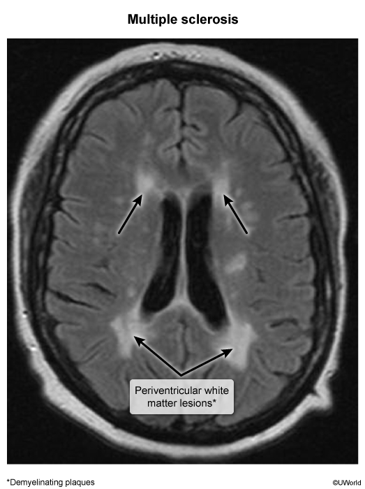
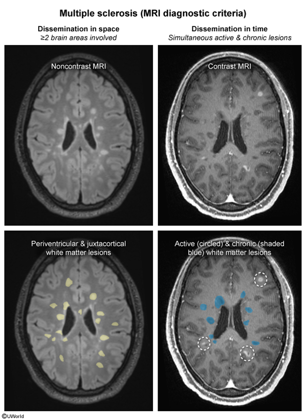
- T1 with Gadolinium: Enhancing lesions indicate acute, active demyelination (provides dissemination in time when combined with non-enhancing older lesions).
- T2/FLAIR: Hyperintense demyelinating plaques in periventricular regions oriented perpendicular to the lateral ventricles (Dawson fingers), corpus callosum, infratentorial regions, and spinal cord.
- Lumbar Puncture / CSF Analysis (used if MRI is non-diagnostic or atypical):
- Oligoclonal bands (OCBs) present on CSF electrophoresis (seen in >85–90% of pts; not present in serum).
- due to increased production of multiple nonspecific IgG sub-fractions in the CSF, which are caused by intrathecal inflammation.
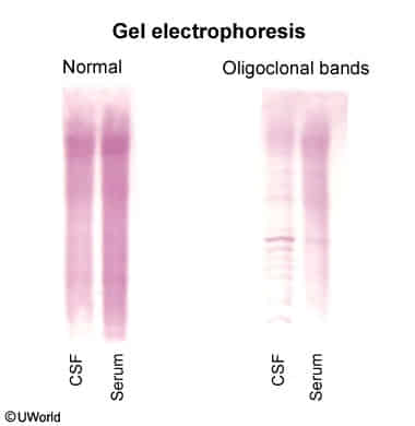
- due to increased production of multiple nonspecific IgG sub-fractions in the CSF, which are caused by intrathecal inflammation.
- Elevated CSF IgG index.
- Normal glucose, normal or mildly elevated protein, mild lymphocytic pleocytosis (<50 WBCs/µL).
- Oligoclonal bands (OCBs) present on CSF electrophoresis (seen in >85–90% of pts; not present in serum).
- Evoked Potentials:
- Visual Evoked Potentials (VEP): Shows delayed P100 latency (indicates subclinical optic nerve demyelination).
Tip
The presence of multiple oligoclonal bands in CSF and their absence in the blood is highly suggestive of MS.
Differential diagnostics
- Transient Ischemic Attack (TIA): c
- Diff by sudden/hyperacute onset (seconds to minutes) restricted to a single vascular territory, rapid resolution (typically <1 hour, always <24 hours), presence of vascular risk factors (HTN, DM2, smoking, AFib), and absence of periventricular demyelinating lesions on MRI. MS relapses evolve subacutely over days to weeks and persist for days to months.
Transverse myelitis
- Etiology & Triggers
- Post-infectious / post-vaccinal immune response (viral URI/GI).
- Demyelinating disease: Multiple Sclerosis (MS), Neuromyelitis Optica Spectrum Disorder (NMOSD [anti-AQP4]), MOGAD. c
- Systemic autoimmune: SLE, Sjögren syndrome, Sarcoidosis.
- Patho
- Autoimmune or post-infectious cross-reactivity (molecular mimicry) triggers an inflammatory cascade targeting the spinal cord.
- Clinical Presentation
- Motor: Bilateral symmetric weakness (paraparesis/tetraparesis); spinal shock (flaccid/areflexic) acutely UMN signs (spasticity, hyperreflexia, (+) Babinski) chronically.
- Sensory: Sharp, discrete sensory level with band-like torso tightness (“corset/belt” sensation); loss of pain, temperature, vibration, and proprioception below lesion. c
- Autonomic: Urinary retention/overflow incontinence, fecal incontinence/constipation, autonomic dysreflexia (if lesion T6).
- Diagnosis
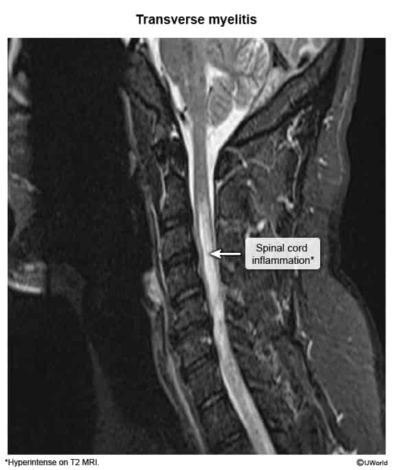
- Spinal MRI w/ contrast (Initial & Best): Shows intramedullary T2 hyperintensity + gadolinium enhancement; rules out compressive cord lesion (e.g., abscess, hematoma, metastasis).
- Brain MRI w/ contrast: Evaluates for demyelinating plaques (MS) or optic nerve lesions (NMOSD).
- Lumbar Puncture (CSF): Lymphocytic pleocytosis, elevated protein, oligoclonal bands (OCB).
- Serologies: Anti-AQP4 IgG (rules in NMOSD), Anti-MOG IgG, ANA, HIV, RPR/VDRL.
- Spinal MRI w/ contrast (Initial & Best): Shows intramedullary T2 hyperintensity + gadolinium enhancement; rules out compressive cord lesion (e.g., abscess, hematoma, metastasis).
- Key Differentials
- Compressive Myelopathy: Extramedullary mass compressing cord on MRI; requires emergent surgical decompression or radiation.
- Anterior Spinal Artery (ASA) Infarction: Hyperacute onset (<minutes to hours); spares dorsal columns (intact vibration & proprioception).
- Guillain-Barré Syndrome (GBS): Ascending paralysis, hyporeflexia, no distinct sensory level, normal MRI, CSF showing albuminocytologic dissociation.
- Multiple Sclerosis (MS): Typically partial/asymmetric cord involvement (<2 vertebral segments), periventricular brain plaques, (+) OCBs.
- NMOSD: Longitudinally extensive transverse myelitis (LETM) (3 vertebral segments), bilateral optic neuritis, (+) anti-AQP4 IgG.
- Management
- 1st-Line (Acute): High-dose IV Methylprednisolone (1 g/day for 3–5 days). c
- 2nd-Line (Steroid-refractory / Severe): Plasma Exchange (PLEX) 5–7 sessions; IVIG as alternative.
- Supportive & Prophylaxis:
- Bladder decompression (Foley catheter / intermittent catheterization).
- DVT prophylaxis (LMWH + SCDs) due to immobility.
- Neuropathic pain (Gabapentin/Pregabalin) & spasticity management (Baclofen).
- Early physical & occupational therapy.
Treatment
- Acute Flares:
- High-dose IV corticosteroids (e.g., methylprednisolone).
- Plasma exchange for severe, steroid-refractory flares.
- Chronic/Disease-Modifying Therapy (DMT):
- Injectables: Interferon-beta, glatiramer acetate.
- Oral: Dimethyl fumarate, fingolimod.
- Infusions (highly effective): Natalizumab (risk of PML due to JC virus), Ocrelizumab (anti-CD20).
- Symptomatic Management:
- Spasticity: Baclofen (GABA agonist), tizanidine (alpha-2 agonist). c
- Neuropathic pain: Gabapentin, pregabalin, TCAs.
- Fatigue: Amantadine, modafinil.
- Urge incontinence: Anticholinergics (e.g., oxybutynin).