Epidemiology
Etiology
Etiologies are broadly categorized as provoked (acute symptomatic) or unprovoked (related to a static or progressive underlying condition).
Common Causes (VITAMINS mnemonic):
- Vascular: Stroke (ischemic or hemorrhagic) is the most common cause in adults, especially the elderly. AVMs.
- Infection: Meningitis, encephalitis, brain abscess, neurocysticercosis (most common cause worldwide).
- Trauma: TBI can cause acute seizures or lead to post-traumatic epilepsy.
- Autoimmune: e.g., Anti-NMDA receptor encephalitis, lupus cerebritis.
- Metabolic: Hypoglycemia, hyponatremia, hypocalcemia, uremia, hepatic encephalopathy.
- Idiopathic/Genetic: Significant portion of childhood-onset epilepsies. Examples include juvenile myoclonic epilepsy, childhood absence epilepsy.
- Neoplasms: Primary or metastatic brain tumors. Seizures can be the presenting symptom.
- Substances: c
- Withdrawal: Alcohol, benzodiazepines, barbiturates.
- Toxicity/Overdose: Cocaine, amphetamines, TCAs, bupropion, tramadol, theophylline.
Etiology by Age
- Neonates (<1 mo): Hypoxic-ischemic encephalopathy (HIE), intracranial hemorrhage, CNS infection, metabolic disturbances (hypoglycemia, hypocalcemia), congenital brain malformations.
- Infants/Children (1 mo - 12 yr): Febrile seizures (most common), genetic epilepsy, CNS infections, trauma, developmental disorders.
- Adolescents (12 - 18 yr): Trauma, genetic epilepsy (e.g., JME), infection, illicit drug use, brain tumors.
- Young Adults (18 - 35 yr): Trauma, alcohol withdrawal, illicit drug use, brain tumors.
- Older Adults (>35 yr): Cerebrovascular disease (stroke), brain tumors, alcohol withdrawal, metabolic disorders (e.g., hypoglycemia).
Seizure Classification
1. Focal (Partial) Onset Seizures
- Originate in one hemisphere of the brain.
- Focal Aware (Simple Partial): Consciousness is fully maintained. Symptoms vary by the affected lobe (e.g., motor, sensory, autonomic, or psychic symptoms like déjà vu). No postictal state.
- Focal Impaired Awareness (Complex Partial): Consciousness is impaired or lost. Often associated with automatisms (e.g., lip-smacking, chewing, hand-wringing). A postictal state (confusion, lethargy) is common. Most commonly arise from the temporal lobe.
- Focal to Bilateral Tonic-Clonic: Starts as a focal seizure and then spreads to involve both hemispheres, becoming a generalized tonic-clonic seizure.
2. Generalized Onset Seizures
- Originate in and rapidly engage bilateral brain networks.
- Tonic-Clonic (Grand Mal): Abrupt loss of consciousness. Tonic phase = stiffening of the body. Clonic phase = rhythmic jerking of limbs. Often with tongue biting, incontinence. Followed by a significant postictal state.
- Absence (Petit Mal): Brief lapse of consciousness (“staring spell”) for 5-10 seconds, often without loss of postural tone. May have subtle motor signs like eyelid fluttering or lip-smacking. No postictal confusion; patient returns immediately to baseline. Classic in children.
- Myoclonic: Sudden, brief, shock-like muscle jerks. Consciousness is usually preserved.
- Atonic: Sudden loss of muscle tone, leading to “drop attacks” and potential injury.
- Tonic: Sustained muscle stiffening without a clonic phase.
| Feature | Absence (Petit Mal) | Focal Impaired Awareness (Complex Partial) |
|---|---|---|
| Patient Age | Child | Usually Adult |
| Key Event | Brief “staring spell" | "Staring spell” + Automatisms (lip-smacking, fumbling) |
| Postictal State | None. Immediately alert. | Present. Confused & drowsy for minutes after. |
| Aura | No | Yes (common; e.g., déjà vu, odd smell, fear) |
| Duration | Short (< 15 seconds) | Longer (1-2 minutes) |
| EEG | Generalized 3 Hz spike-wave | Focal spikes (e.g., temporal lobe) |
| 1st Line Tx | Ethosuximide | Lamotrigine, Levetiracetam |
Clinical features
Diagnostics
- Initial / Screening Evaluation:
- Fingerstick glucose (rule out immediate hypoglycemia).
- Key Labs: BMP (Na, Ca, Mg, BUN/Cr), CBC, toxicology screen, UPT (all females of childbearing age). c
- Transient Post-Ictal Labs:
- Elevated serum prolactin (drawn within 10–20 min; helps differentiate true GTC from PNES).
- Elevated serum lactic acid (anion gap metabolic acidosis due to intense muscle contraction; self-resolves within 60–90 min).
- Elevated CK (rhabdomyolysis).
- Neuroimaging:
- Non-contrast Head CT: Initial emergency imaging to rule out acute intracranial hemorrhage, mass effect, or stroke.
- Brain MRI with seizure protocol: Modality of choice for evaluating non-emergent or first-time unprovoked seizures (detects mesial temporal sclerosis, low-grade tumors, cortical dysplasias, vascular malformations).
- Confirmatory / Gold Standard:
- Electroencephalography (EEG): Essential for classifying seizure type, locating epileptogenic focus, and identifying syndromes.
- Absence: 3-Hz spike-and-wave discharges.
- JME: Generalized 4–6 Hz polyspike-and-wave discharges.
- Herpes Encephalitis: Periodic lateralized epileptiform discharges (PLEDs) over temporal lobes.
- Continuous Video-EEG Monitoring: Gold standard for diagnosing psychogenic non-epileptic seizures (PNES) and pre-surgical evaluation for refractory epilepsy.
- Electroencephalography (EEG): Essential for classifying seizure type, locating epileptogenic focus, and identifying syndromes.
- Lumbar Puncture (LP): Indicated only if clinical suspicion of CNS infection (fever, meningismus, altered mental status) or in immunocompromised patients, performed after ruling out mass effect with CT.
Differential Diagnostics
- Breath-Holding Spells (Cyanotic vs Pallid):
- Diff: Typical age 6 months to 2 years (resolves by age 5); always provoked by emotional distress, anger, frustration, or minor pain/trauma; crying followed by breath-holding in expiration cyanosis or severe pallor LOC brief clonic jerks/twitching; no true post-ictal confusion (immediate rapid recovery to baseline); normal EEG. Strongly associated with iron deficiency anemia (check CBC and serum ferritin; iron supplementation reduces episode frequency). c
- Psychogenic Non-Epileptic Seizures (PNES):
- Diff: Asynchronous limb thrashing, pelvic thrusting, side-to-side head shaking, forced eyelid closure with resistance to manual opening, preserved pupillary light reflex, absence of post-ictal lactic acidosis/prolactin rise; normal EEG during event.
Treatment
Acute management
- Early status epilepticus (5–20 minutes): first-line therapy
- IV benzodiazepine (lorazepam OR diazepam)
- Persistent status epilepticus (20–40 minutes): second-line therapy
- IV fosphenytoin
- IV valproic acid
- IV levetiracetam
- Refractory status epilepticus (40–60 minutes)
- Options include repeat second-line therapy or induction of coma (e.g., with IV propofol, thiopental, midazolam, or pentobarbital).
Long-term management
- Focal
- Lamotrigine
- Levetiracetam
- Phenytoin
- Carbamazepine
- Oxcarbazepine
- Phenobarbital (children)
- Generalized
Complications
Acute
- Postictal lactic acidosis: postictal transient anion gap metabolic acidosis with increased lactic acid and reduced serum bicarbonate (usually resolves spontaneously within 60–90 minutes after seizure activity stops) c
- Lactic acidosis results from excess muscle exertion in the setting of inadequate oxygen delivery.
Status epilepticus
- Definition & Timing:
- Seizure ≥5 min or ≥2 discrete seizures without return to neurologic baseline.
- t₁ (5 min): start emergency pharmacotherapy immediately; do not wait 30 min.
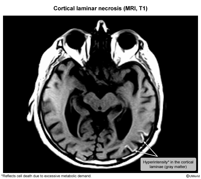
- t₂ (30 min): mark of irreversible excitotoxic neuronal injury.
- Etiology & Risk Factors:
- ASM nonadherence (most common in known epilepsy).
- Acute CNS insults: ischemic stroke, ICH/SAH, meningitis/encephalitis, TBI, mass lesion.
- Metabolic/Toxic: hypoglycemia, hyponatremia, hypocalcemia, alcohol/BZD withdrawal, INH toxicity, cocaine.
- Special: Eclampsia in pregnancy/postpartum.
- Clinical Pearls & Diagnostics:
- Nonconvulsive SE (NCSE): suspect if pt has persistent AMS/coma and fails to awaken after motor convulsions cease.
- First bedside test: Finger-stick glucose (treat hypoglycemia immediately).
- Diagnostic gold standard: Continuous EEG (cEEG) (required to diagnose NCSE and titrate sedation in refractory SE).
- Etiologic workup (parallel to tx): CMP (Na, Ca, renal/hepatic fxn), tox screen, ASM serum levels, noncontrast head CT (post-stabilization), LP (if CNS infection suspected).
- Stepwise Management:
- 0–5 min (Stabilization & ABCs):
- Position pt, suction airway, high-flow O₂, continuous pulse ox/telemetry, obtain IV/IO access.
- Give IV Dextrose (if hypoglycemic) + thiamine (if alcohol use disorder).
- 5–10 min (First-line: Benzodiazepines):
- IV Lorazepam (0.1 mg/kg, max 4 mg); may repeat once.
- IM Midazolam (10 mg if >40 kg, 5 mg if 13–40 kg) if no IV access.
- IV Diazepam (0.15–0.2 mg/kg, max 10 mg) alternative.
- 10–20 min (Second-line: Non-BZD IV ASMs):
- IV Levetiracetam (60 mg/kg, max 4,500 mg) — fewest drug interactions. c
- IV Fosphenytoin (20 mg PE/kg, max 1,500 mg PE) — requires cardiac monitoring (arrhythmia/hypotension risk).
- IV Valproate (40 mg/kg, max 3,000 mg) — avoid in pregnancy/hepatic failure.
- 20–40+ min (Refractory SE):
- Endotracheal intubation + ICU admission + urgent cEEG. c
- Continuous anesthetic infusion: Propofol, Midazolam, or Pentobarbital/Thiopental.
- Titrate to electrographic seizure cessation or burst suppression.
- Etiology-Specific Antidotes:
- Eclampsia → IV Magnesium Sulfate (MgSO₄) + delivery.
- Isoniazid (INH) overdose → IV Pyridoxine (Vitamin B₆).
- Severe symptomatic hyponatremia → 3% Hypertonic saline.
- 0–5 min (Stabilization & ABCs):
- Differential Diagnosis:
- Psychogenic Non-Epileptic Seizures (PNES): forced eye closure, pelvic thrusting, out-of-phase limb movements, normal EEG.
- Postictal State vs NCSE: postictal shows gradual clinical recovery; NCSE has persistent AMS/coma and requires EEG to confirm.
- Convulsive Syncope: brief myoclonic jerks (<15 sec) following prodrome, immediate return to baseline orientation.
- Complications:
- Lactic acidosis, rhabdomyolysis, hyperkalemia, acute kidney injury (AKI).
- Aspiration pneumonia, neurogenic pulmonary edema, hyperthermia.
- Permanent cognitive/neurologic deficits, propofol infusion syndrome (PRIS) with prolonged high-dose propofol.