↑ ICP → ↓ CPP → compensatory activation of the sympathetic nervous system → ↑ systolic blood pressure → stimulation of aortic arch baroreceptors → activation of the parasympathetic nervous system (vagus) → bradycardia c
↑ Pressure on brainstem → dysfunction of respiratory center → irregular breathing
Clinical features
General ↑ ICP:
Headache (worse in AM, worse supine, worsened by Valsalva/coughing).
Nausea/Vomiting (often projectile, without nausea).
Papilledema (blurring of optic disc margins).
Altered Mental Status (AMS), depressed level of consciousness.
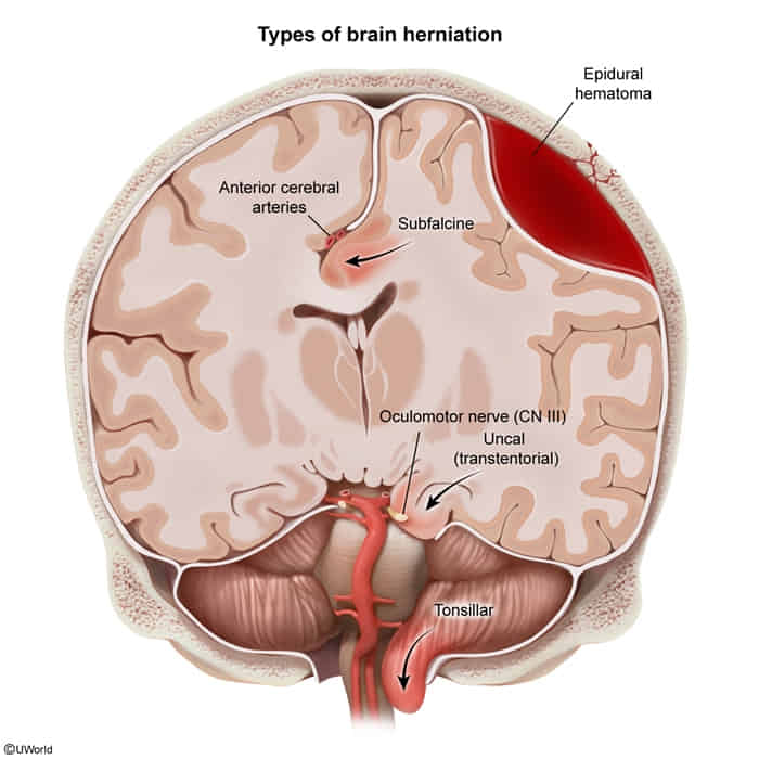
What: Cingulate gyrus is displaced under the falx cerebri. This is the most common type of herniation.
Vessels: Compression of the anterior cerebral artery (ACA) can occur.
Clinical signs: May be asymptomatic or cause contralateral lower limb weakness due to motor cortex ischemia.
Transtentorial (Uncal)
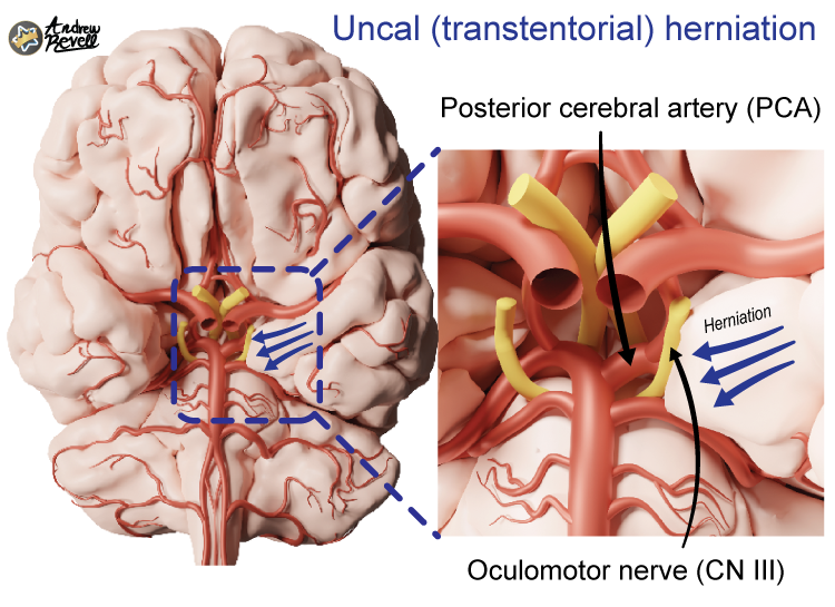
What: Medial temporal lobe (specifically the uncus) is forced through the tentorial notch.
CN III Palsy: This is a classic and critical finding.
Early: Compression of parasympathetic fibers on the exterior of CN III causes an ipsilateral fixed and dilated pupil (“blown pupil”).
Late: Compression of motor fibers of CN III leads to a “down and out” gaze.
Vessels: Ipsilateral posterior cerebral artery (PCA) compression can cause contralateral homonymous hemianopsia.
Motor: Compression of the contralateral cerebral peduncle against the tentorium (Kernohan’s Notch) causes ipsilateral hemiparesis (a false-localizing sign).
Brainstem: Brainstem displacement can cause tearing of paramedian basilar artery branches, leading to fatal Duret hemorrhages.
Central Herniation
What: Downward displacement of the diencephalon and brainstem through the tentorial notch.
Clinical signs: Leads to a rapid decline in consciousness, decorticate posturing progressing to decerebrate posturing, and eventually death.
Tonsillar
What: Cerebellar tonsils are forced through the foramen magnum.
Clinical signs: Compresses the medulla, which contains the cardiorespiratory centers. This leads to respiratory arrest, circulatory collapse, and rapid death.
Clinical pearl: This is why a lumbar puncture (LP) is contraindicated in patients with suspected ↑ICP until an intracranial mass lesion has been ruled out by imaging (e.g., CT scan). An LP can create a pressure gradient that precipitates tonsillar herniation.
Airway & Breathing: Intubate if GCS ≤ 8; maintain normoxia (PaO2 > 60 mmHg) and normocapnia (PaCO2 35–40 mmHg).
Hemodynamic Control: Maintain CPP = 60–70 mmHg (CPP = MAP – ICP); avoid hypotension (SBP < 90 mmHg). Use IV vasopressors (e.g., norepinephrine) if needed to support MAP.
Positioning: Elevate head of bed (HOB) to 30–45° with neck midline to facilitate jugular venous outflow.
Analgesia/Sedation: IV propofol or fentanyl to prevent coughing, agitation, and reduce cerebral metabolic demand (CMRO2).
Temperature Control: Aggressive antipyretics and cooling (fever increases CMRO2 and ICP).
2. Medical Interventions (Tier 2):
Osmotic Therapy:
Hypertonic Saline (3% or 23.4% NaCl): First-line in hypotensive/hypovolemic pts; draws water from parenchyma into vascular space. Target serum Na 145–155 mEq/L. c
Mannitol (20%): Bolus 0.5–1.0 g/kg IV. Contraindicated in hypotension, hypovolemia, and severe renal failure. Target serum Osm < 320 mOsm/kg.
Glucocorticoids (Dexamethasone):
Indicated only for vasogenic edema due to CNS tumors/metastases or abscesses.
Contraindicated in TBI, stroke, and ICH (worsens mortality/outcomes).
Hyperventilation (Target PaCO2 26–30 mmHg):
Causes cerebral vasoconstriction -> rapid reduction in cerebral blood volume.
CSF Drainage: External Ventricular Drain (EVD) for therapeutic CSF removal and continuous monitoring.
Surgical Evacuation / Decompression: Urgent craniotomy/craniectomy for hematoma evacuation or decompressive hemicraniectomy for refractory malignant edema/stroke.
Metabolic Suppression: Barbiturate coma (Pentobarbital) or therapeutic hypothermia (32–35°C) to suppress CMRO2 in medically refractory cases without surgical targets.