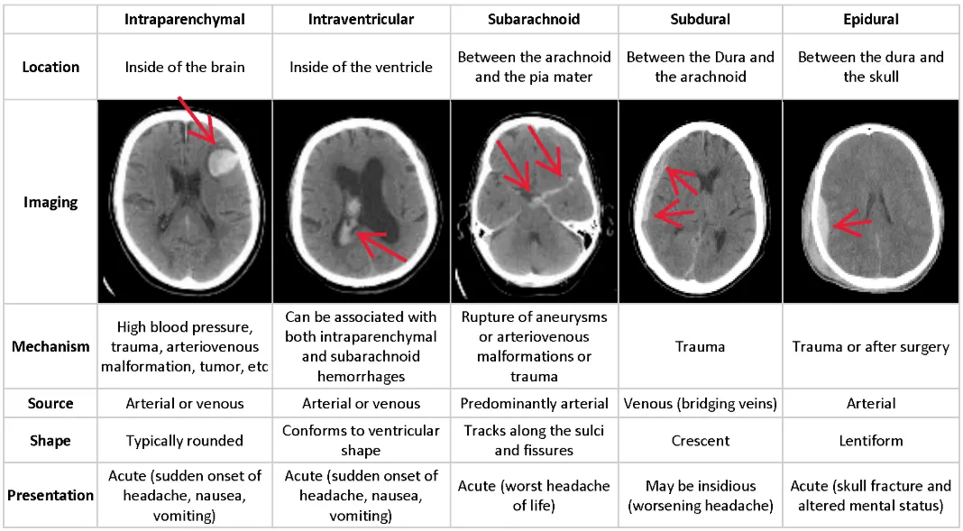
| Type | Vessel / Cause | Key Associations | CT Appearance |
|---|---|---|---|
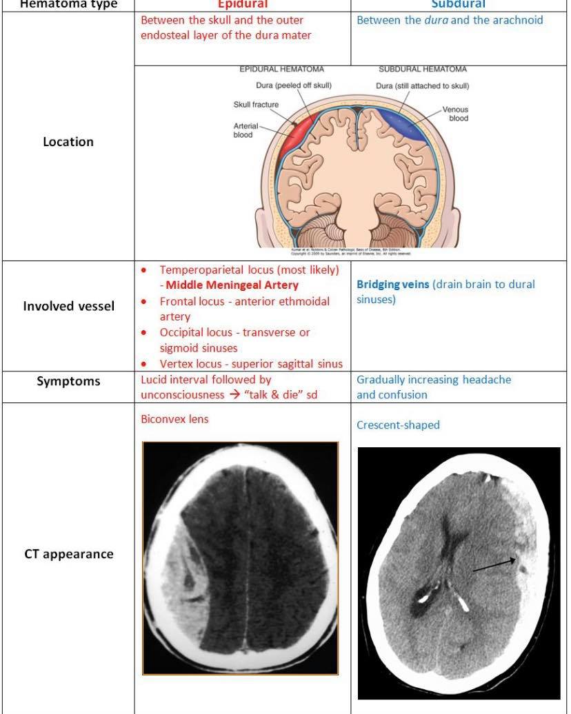
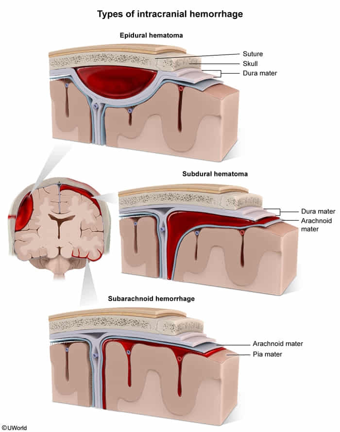
| Epidural | Middle Meningeal Artery | Pterion fracture, Lucid interval | Lens-shaped (Biconvex) Stops at sutures |
| Subdural | Bridging Veins c | Elderly, Alcoholics, Shaken Baby | Crescent-shaped Crosses sutures |
| Subarachnoid | Berry Aneurysm / AVM | ”Thunderclap” headache, Meningeal signs | Blood in Sulci LP: Xanthochromia |
| Intraparenchymal | Charcot-Bouchard (HTN) | HTN, Amyloid angiopathy, Basal Ganglia | Focal hyperdensity within brain |
| Intraventricular | Germinal Matrix | Premature infants (<32 wks) | Blood inside Ventricles |
Epidemiology
Etiology
- Chronic HTN (most common overall): Causes Charcot-Bouchard microaneurysms in deep penetrating lenticulostriate arteries (basal ganglia, thalamus, pons). c
- Cerebral Amyloid Angiopathy (CAA): Most common cause of spontaneous lobar/cortical ICH in the elderly; associated with Alzheimer disease. c
- Anticoagulant or thrombolytic therapy.
- Sympathomimetic illicit drugs (e.g., cocaine, amphetamines). c
- Vascular malformations (AVMs, cavernous hemangiomas) in younger pts.
- Common locations: Putamen/Internal capsule (most common, ~50%), Thalamus, Pons, Cerebellum, Subcortical white matter (Lobar).
Pathophysiology
- Nontraumatic mechanisms of hemorrhage
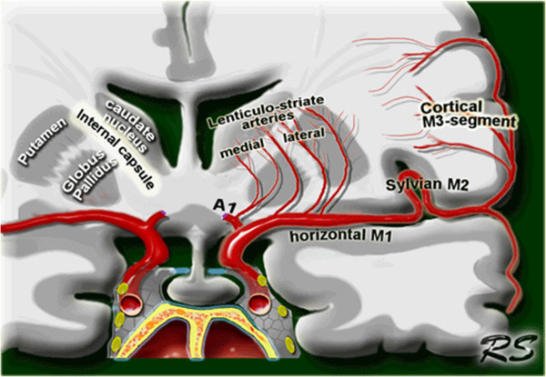
- Chronic arterial hypertension → lipohyalinosis of lenticulostriate vessels (which supply the basal ganglia) and/or formation and rupture of Charcot-Bouchard microaneurysms → lacunar strokes (ischemia) of the basal ganglia. See also Ischemic stroke > Lacunar infarction
- Putamen most commonly affected, which is supplied by lenticulostriate arteries
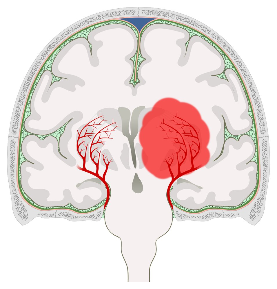
Charcot-Bouchard aneurysms vs Saccular (berry) aneurysms
Feature Charcot-Bouchard Saccular (Berry) Etiology Chronic HTN Congenital weakness + Hemodynamics Pathology Lipohyalinosis of microvessels Lacking Internal Elastic Lamina & Media Location Deep Brain (Basal Ganglia, Thalamus) Circle of Willis Bifurcations (ACom > PCom) Vessels Lenticulostriate arteries Medium-sized arteries Rupture Intraparenchymal Hemorrhage Subarachnoid Hemorrhage Symptoms Focal deficits (Hemiparesis) “Thunderclap” Headache, Meningismus Associations Lacunar strokes ADPKD, Ehlers-Danlos, Marfan -aneurysms/../../../appendix/pasted-image-20250304102040.png)
-aneurysms/../../../appendix/pasted-image-20240526113641.png)
- Saccular: This term means “resembling a sac.” Saccular aneurysms are outpouchings or bulges on one side of a blood vessel wall.
- Berry: The “berry” description refers to the characteristic round shape of these aneurysms. They look like a berry connected to the main artery.
- Other locations: thalamus (second most common) and infratentorial parts of the brain (e.g., pons, cerebellum)
- Putamen most commonly affected, which is supplied by lenticulostriate arteries
- Cerebral amyloid angiopathy: deposition of β-amyloid peptides in vessel walls → focal damage with formation of microaneurysms → rupture → recurrent lobar intracerebral hemorrhage
- Chronic arterial hypertension → lipohyalinosis of lenticulostriate vessels (which supply the basal ganglia) and/or formation and rupture of Charcot-Bouchard microaneurysms → lacunar strokes (ischemia) of the basal ganglia. See also Ischemic stroke > Lacunar infarction
- Traumatic: blunt or penetrating injury → damage to vessels
Clinical features
- Headache
- Focal neurologic signs and symptoms
- Putaminal hemorrhage: contralateral hemiparesis or hemiplegia with less severe contralateral hemisensory loss; eyes deviate toward the side of the hematoma
- Thalamic hemorrhage: contralateral hemiparesis, contralateral hemisensory loss, decreased consciousness, wrong way eyes
- Putaminal hemorrhage: contralateral hemiparesis or hemiplegia with less severe contralateral hemisensory loss; eyes deviate toward the side of the hematoma
Mnemonic
- The dorsal striatum is composed of both the caudate and the putamen.
- The caudate is involved in both motor activity as well as more cognitive functions, while the putamen is primarily involved in motor activity.
- The caudate nucleus is the most caugnitive part of the basal ganglia.
- When you see put-am-en on an exam, “put-an-em” for Motor.
- Course
- Symptoms typically progress gradually over minutes to a few hours
- Focal deficits worsen with expansion of the hematoma
- Late: symptoms of increased ICP
- Nausea and vomiting
- Confusion and loss of consciousness
- Bradycardia
- Fixed pupils
Diagnostics
- Initial & Best Step: Non-contrast Head CT
- Identifies acute hyperdense (bright white) parenchymal collection with surrounding hypodense edema/mass effect.
- Key Labs:
- Coagulation panel (PT/INR, PTT, Platelets): To identify coagulopathy.
- Toxicology screen: Rule out sympathomimetic drug use. c
- CBC, BMP, Type & Screen.
- Secondary/Confirmatory Vascular Imaging:
- CTA or MRI/MRA brain: Performed once stable if suspecting secondary causes (AVM, aneurysm, venous sinus thrombosis, hemorrhagic tumor).
Treatment
- 1. Airway/ABCs: Intubate if GCS ≤ 8; elevate HOB 30°.
- 2. BP Control (Immediate): Target SBP 130–140 mmHg with IV Nicardipine or Labetalol.
- 3. Reverse Anticoagulation (Stat):
- Warfarin: 4F-PCC + IV Vitamin K.
- Heparin: Protamine sulfate.
- Dabigatran: Idarucizumab.
- Factor Xa Inhibitors (Apixaban, Rivaroxaban): Andexanet alfa or 4F-PCC.
- 4. Manage ↑ ICP: IV Mannitol or 3% Hypertonic Saline.
- 5. Neurosurgery:
- Cerebellar hematoma ≥ 3 cm or brainstem compression Emergent surgical decompression.
- EVD placement for acute obstructive hydrocephalus.