Epidemiology
Etiology
Risk factors
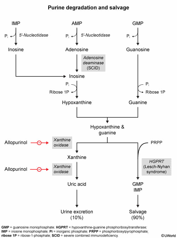
- Increased uric acid production
- Dietary sources
- Purine-rich foods (eg, seafood, red meat)
- Fructose-containing & alcoholic beverages (particularly beer)
- High cell turnover: Myeloproliferative disorders, leukemia/lymphoma, Tumor Lysis Syndrome, psoriasis. c
- Lesch-Nyhan syndrome (deficiency of HGPRT)
- ↑ Phosphoribosyl pyrophosphate activity
- Dietary sources
- Decreased uric acid clearance
- Chronic kidney disease
- Volume depletion
- Diuretics (eg, thiazide)
- Niacin
- Cyclosporine & tacrolimus
- Rapid decline in uric acid levels
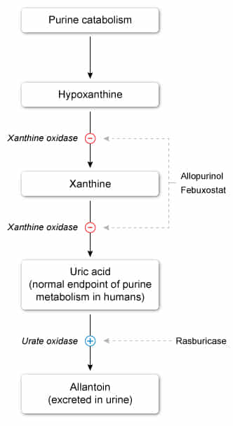
- Xanthine oxidase inhibitors (eg, allopurinol)
- Uricosuric drugs (eg, probenecid)
Pathophysiology
- Gout
- An inflammatory crystal arthropathy that is caused by the precipitation and deposition of uric acid crystals in synovial fluid and tissues.
- It is typically associated with hyperuricemia, but can also occur if uric acid levels are normal.
- Uric acid
- An end-product of purine metabolism that is excreted by the kidneys
- Has somewhat poor water solubility
- Predisposes to gout
- Triggers of urate crystal deposition
- ↑ Uric acid levels (due to insufficient excretion or increased production of purines)
- Acidosis
- Low temperature (e.g., cool peripheral joints)
- Crystalline arthritis: supersaturation of uric acid in extracellular fluid → intraarticular uric crystal precipitation (coated by IgGs) → phagocytosis by polymorphonuclear cells → release of inflammatory mediators and enzymes → local joint inflammation
- Chronic effects: repeated attacks → aggregations of urate crystals and giant cells (tophi) → deformities and arthritis
Clinical features
- Acute Gouty Arthritis:
- Sudden, excruciating monoarticular joint pain, erythema, warmth, and marked swelling.
- Peaks within 12–24 hours; often begins overnight or early morning.
- Podagra: 1st metatarsophalangeal (MTP) joint is most common (> 50% initial flares); also affects midfoot, ankles, knees, wrists.
- Triggers: Trauma, surgery, large meal, alcohol binge, dehydration, initiation of urate-lowering therapy (ULT).
- Chronic Tophaceous Gout:
- Develops after years of untreated/poorly controlled disease.
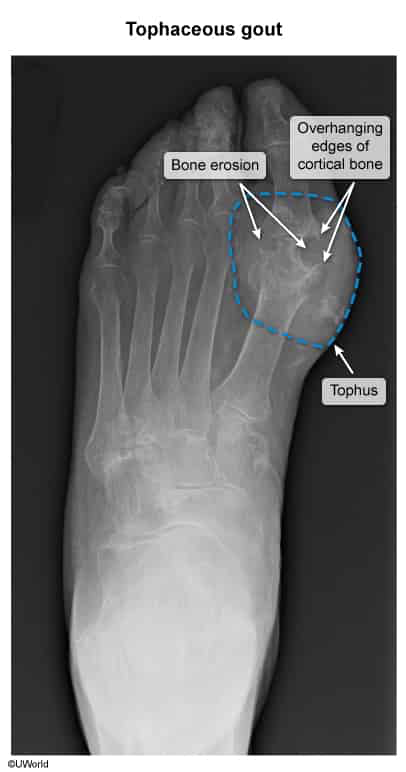
- Tophi: Painless or tender nodular chalky deposits of MSU crystals in soft tissues, tendons, and cartilage (helix of ear, olecranon bursa, Achilles tendon).
- Can lead to chronic joint deformities mimicking rheumatoid arthritis (RA).
Diagnostics
- Initial & Confirmatory / Gold Standard: Arthrocentesis with Synovial Fluid Analysis.
- Polarized Light Microscopy: Needle-shaped, negatively birefringent crystals (yellow when parallel to polarized axis, blue when perpendicular).
- Cell Count: Inflammatory (WBC 2,000–50,000/mm³, occasionally > 50,000/mm³) with PMN predominance (> 50%).
- Gout: >2,000/mm3
- Septic arthritis: > 50,000/mm3
- Gram Stain & Culture: Mandatory to rule out concomitant septic arthritis.
- Key Labs:
- Serum Uric Acid: Usually elevated, but can be normal or low during an acute flare (urate acts as an acute-phase reactant and drops into tissues). Do not use serum urate alone to diagnose or rule out an acute attack.
- ↑ ESR, ↑ CRP, and peripheral leukocytosis during acute attacks.
- Imaging:
- Plain Radiographs (X-ray):
- Acute: Soft tissue swelling only.
- Chronic: “Punched-out” lytic bone erosions with sclerotic margins and overhanging edges (“rat-bite” lesions), preservation of joint space until late.
- Ultrasound / DECT: “Double contour sign” on articular cartilage; useful if arthrocentesis is inconclusive or inaccessible.
- Plain Radiographs (X-ray):
Treatment
Tip
- NSAIDs (e.g., naproxen, indomethacin) preferred if no contraindications
- Colchicine used as second-line therapy
Acute gout flare
NSAIDs
- Naproxen or an alternative (e.g., indomethacin, ibuprofen)
- Contraindicated in PUD
Colchicine
- Mechanism of action: binds and stabilizes tubulin subunits → inhibits microtubule polymerization → inhibits phagocytosis of urate crystals, neutrophil activation, migration, and degranulation
- Adverse effects
- Gastrointestinal symptoms, e.g., diarrhea, nausea, vomiting, and abdominal pain, are the most common.
- Rhabdomyolysis , myopathy
- Polyneuropathy
- Cardiac toxicity, arrhythmias
- Nephrotoxicity
- Myelosuppression
- CNS symptoms (e.g., fatigue, headache)
Tip
Chronic gout
- Urate-lowering therapy (ULT) is recommended for chronic gout.
- First-line: xanthine-oxidase inhibitors (allopurinol)
- Second-line: uricosurics (probenecid)
- Third-line: recombinant uricase (pegloticase, rasburicase)
- Also used in Tumor lysis syndrome
- Administer anti-inflammatory prophylaxis before initiating ULT as ULT may trigger, prolong, or worsen an acute gout flare.
- Lowering of serum urate levels likely causes preexisting urate crystal deposits to dissolve and become mobile.
Uricosurics
- Probenecid
- Mechanism of action
- Inhibition of uric acid reabsorption along renal proximal convoluted tubules → increased renal elimination
- Side effects
- Nephrolithiasis > Uric acid stones
- Hypersensitivity reactions (sulfa allergy due to being a sulfonamide derivative)