Tip
- Hydatidiform mole: need sperm + oocyte.
- Teratoma: just oocyte.
Epidemiology
Etiology
Tip
The terms “partial” and “complete” refer to the extent of abnormal tissue growth and the presence or absence of fetal tissue. In a complete mole, no normal tissue is present, whereas in a partial mole, there may be some but it’s still non-viable.
- Complete Mole
- Karyotype: 46,XX (most common) or 46,XY.
- Mechanism: Empty egg (no maternal DNA) + 1 sperm (duplicates DNA) OR + 2 sperm. All genetic material is paternal (Androgenesis). t
- Fetal Parts: Absent.
- Uterine Size: > Dates (enlarged).
- -hCG: Extremely high (>100,000 mIU/mL).
- Risk of Malignancy: 15–20% (Gestational Trophoblastic Neoplasia/Choriocarcinoma).
- Immunostain: p57 negative (p57 is maternally expressed; no maternal DNA = no expression).
- Partial Mole
- Karyotype: 69,XXX, 69,XXY, or 69,XYY.
- Mechanism: Normal egg + 2 sperm (or 1 sperm that duplicates).
- Fetal Parts: Present (fetal tissue often seen).
- Uterine Size: Normal or Small for dates.
- -hCG: Normal or slightly elevated.
- Risk of Malignancy: Low (<5%).
- Immunostain: p57 positive (contains maternal DNA).
Pathophysiology
Clinical features
- Complete Mole:
- Painless vaginal bleeding (dark red/brown “prune juice” discharge) at 6-16 wks GA.
- Uterine size larger than expected for GA.
- Hyperemesis gravidarum (due to massively ↑ β-hCG).
- Early-onset preeclampsia with severe features (< 20 wks GA).
- Hyperthyroidism (heat intolerance, tachycardia; β-hCG cross-reacts with TSH receptors). c
- Bilateral theca lutein cysts (ovarian enlargement/pelvic pressure from LH-like stimulation).
- Partial Mole:
- Often presents as missed or incomplete abortion.
- Mild vaginal bleeding, pelvic pain.
- Uterine size normal or small for GA.
Diagnostics
DDx
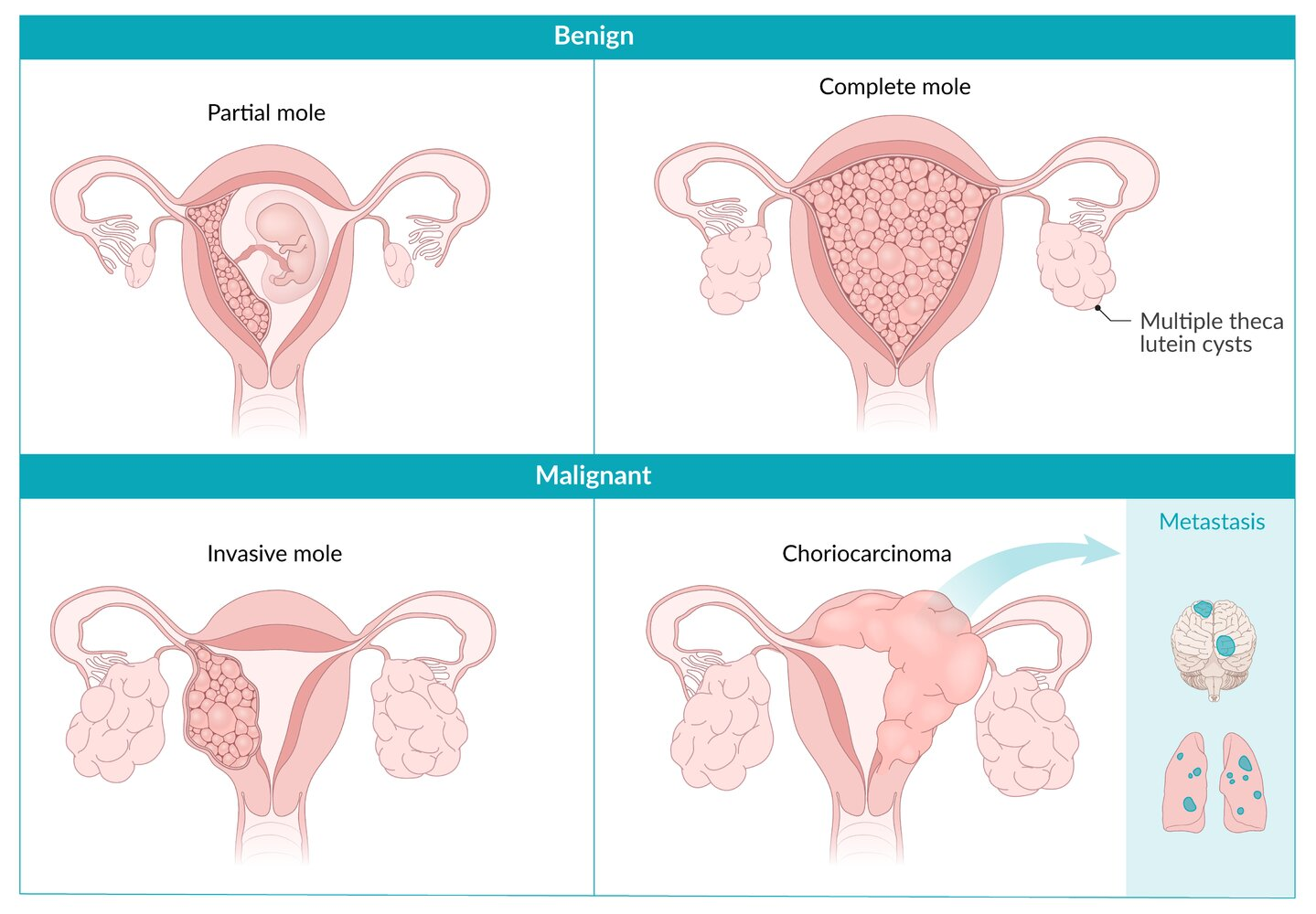
Comparison of choriocarcinoma, hydatidiform mole, and teratoma
Link to original
Feature Choriocarcinoma Hydatidiform Mole Teratoma Nature Malignant Premalignant Benign (usually) Origin Trophoblasts (often from molar pregnancy) Trophoblasts (from abnormal fertilization) Germ cells Karyotype Aneuploid (often derived from mole) 46,XX (Complete) or 69,XXX/XXY (Partial) 46,XX Key Histo Anaplastic trophoblasts, NO villi Hydropic (swollen) villi Mature tissue (hair, teeth, etc.) β-hCG Massively ↑ (>100k) Massively ↑ (>100k, Complete) Normal Key Sx Lung mets (hemoptysis, dyspnea) Uterine size > dates, preeclampsia <20wks Asymptomatic or ovarian torsion Ultrasound Solid uterine mass ”Snowstorm” appearance (Complete) Cyst with calcifications/fat Treatment Chemotherapy (Methotrexate) Suction Curettage Surgical Removal (Cystectomy)
Treatment
- Immediate / First-Line:
- Suction dilation and curettage (D&C) under US guidance (treatment of choice for uterine evacuation regardless of size).
- Give anti-D immune globulin (RhoGAM) to Rh-negative mothers immediately.
- Treat systemic manifestations (e.g., β-blockers for symptomatic thyrotoxicosis; antihypertensives/magnesium sulfate for preeclampsia).
- Definitive / Alternative (Surgical):
- Hysterectomy (preserves ovaries): Indicated if childbearing is complete or in older women (>40 years) to decrease malignant sequelae.
- Post-Evacuation Surveillance (Crucial):
- Monitor serial quantitative serum β-hCG weekly until undetectable for 3 consecutive weeks, then monthly for 6 months.
- Strict contraception (OCPs, implants; avoid IUD until β-hCG is negative due to perforation risk) during the entire 6-month monitoring window to prevent confounding rises in β-hCG.
- Refractory / Gestational Trophoblastic Neoplasia (GTN):
- Diagnosed if β-hCG plateaus (4 values over 3 weeks) or rises (3 values over 2 weeks), or if histologic choriocarcinoma is present.
- Chemotherapy: Single-agent Methotrexate or Actinomycin D (low-risk GTN); multi-agent EMA-CO (Etoposide, Methotrexate, Actinomycin D, Cyclophosphamide, Vincristine) for high-risk/metastatic disease.