Shigellosis
Etiology: Shigella spp. Gram-negative, non-motile, non-lactose fermenting, H2S-negative bacilli.
S. sonnei : Most common in US/developed countries.S. flexneri : Most common in developing countries.S. dysenteriae : Causes severe disease/outbreaks (produces Shiga toxin ).
Transmission: Fecal-oral route, contaminated food/water, person-to-person contact.
High infectivity: Very low infectious dose (10–100 organisms); highly resistant to gastric acid.
Risk factors: Daycare centers, crowded living conditions, international travel, MSM , poor sanitation.
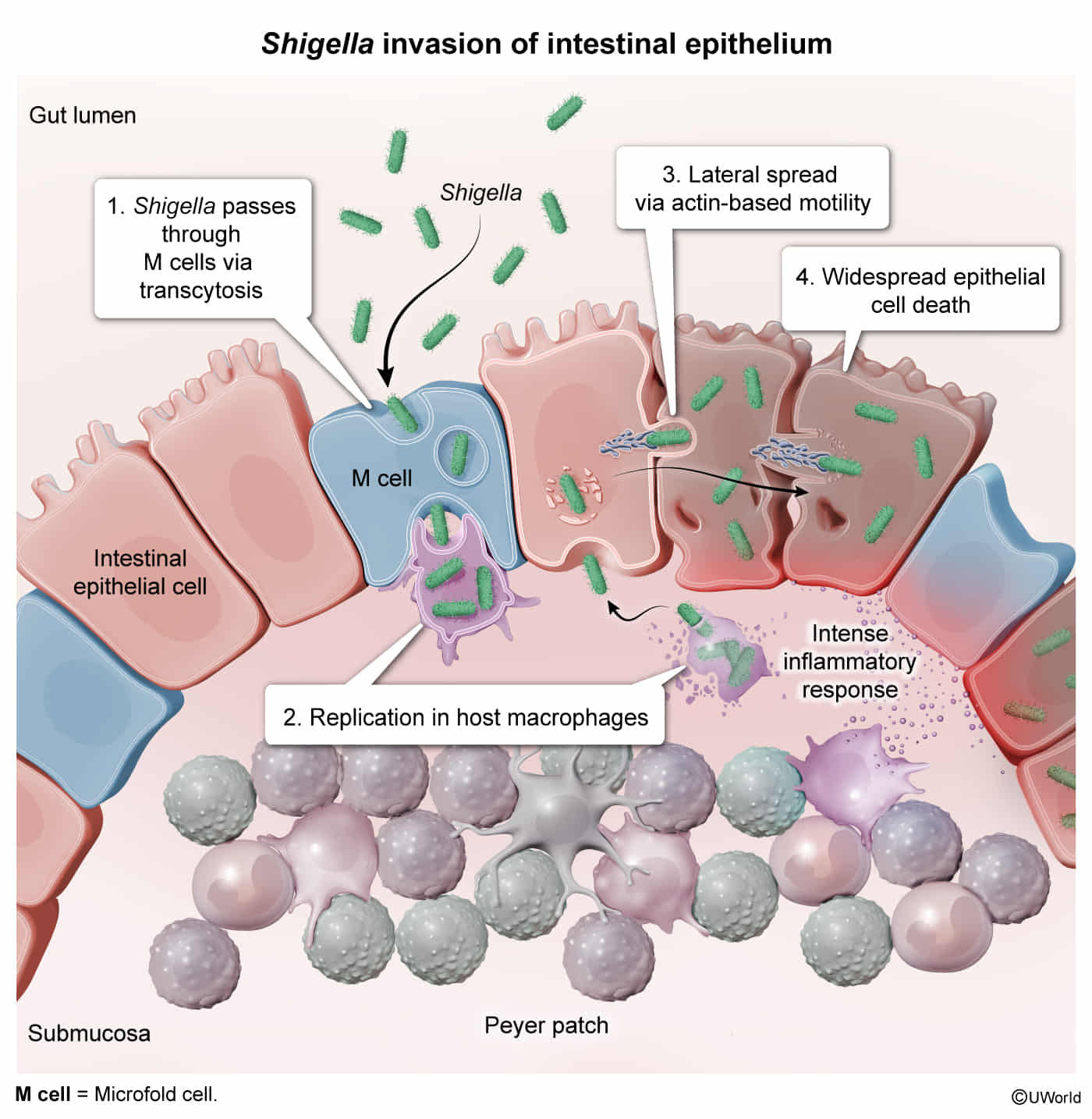
Mechanism: Highly virulent with a very low infectious dose (as few as 10-100 organisms) because it is acid-stable. It invades the gastrointestinal mucosa, primarily in the large intestine, through M cells of Peyer’s patches . It then escapes the phagosome and spreads laterally from cell to cell using the host’s actin filaments (no hematogenous spread) , causing mucosal ulceration and inflammation.
Toxin: Some strains, particularly S. dysenteriae , produce Shiga toxin, which inactivates the 60S ribosomal subunit, inhibiting protein synthesis. However, the primary cause of pathology is direct mucosal invasion.
Clinical features
Incubation: 1–4 days.
Initial phase : High fever, crampy abdominal pain, watery diarrhea . c Classic presentation: Progression to frequent, small-volume bloody/mucoid diarrhea (dysentery) with severe tenesmus (painful urge to defecate).
PE: Lower abdominal tenderness, hyperactive bowel sounds, dehydration signs.
Pediatric feature: High risk of febrile seizures
Cholera
Pathogen: Vibrio cholerae
Infectivity
Acid-labile (grows well in an alkaline medium) High infective dose required (over 108 pathogens)
Gastric acid provides a natural barrier against V. cholerae infection
Patients on long PPI are more easily to get infected t
Noncholera Vibrio infection
Transmission / Exposure :
Ingestion of raw or undercooked seafood (especially raw oysters ).
Direct exposure of open wounds/cuts to contaminated saltwater or brackish water or marine life.
High-Risk Populations (V. vulnificus bacteremia/severe wound infection):
Chronic Liver Disease (CLD) / Cirrhosis Hemochromatosis c Immunocompromised states (DM, ESRD, HIV, active malignancy).
Clinical Presentation :
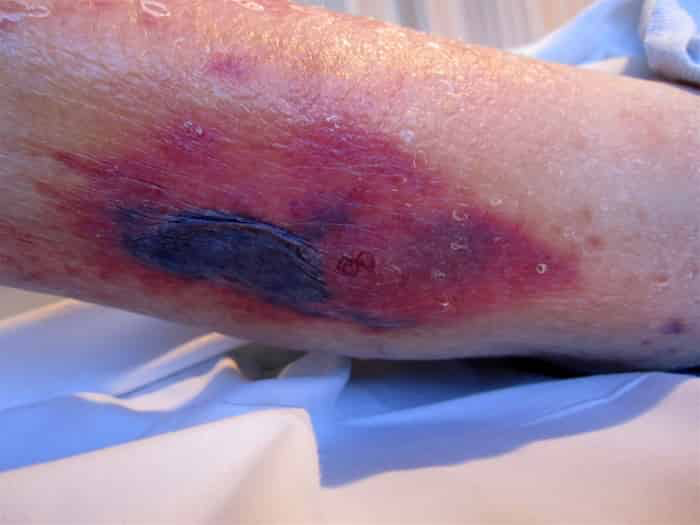
V. vulnificus Rapid (within hours) necrotizing soft-tissue infection w/ hemorrhagic bullae ; fulminant septic shock. c V. parahaemolyticus
Diagnosis : Blood/wound culture on TCBS agar (Gram-negative curved rods).Key Differential :
Aeromonas hydrophila freshwater or leeches (vs saltwater/marine environments for Vibrio ).Streptococcus pyogenes (GAS Necrotizing Fasciitis)Clostridium perfringens (Gas Gangrene)crepitus , and Gram-positive rods on stain.
Management :
V. vulnificus / SepsisEmergent surgical debridement + IV Doxycycline + Ceftriaxone .Gastroenteritis : Supportive (oral rehydration).
Complications : Septic shock, DIC, necrotizing fasciitis (>50% mortality in cirrhotic pts).
Campylobacter enteritis (campylobacteriosis)
Epidemiology & Risk Factors
Pathogen : Campylobacter jejuni (curved/seagull-shaped GNR, thermophilic [42°C], oxidase [+]).#1 cause of bacterial gastroenteritis in US.Source : Undercooked poultry (most common), unpasteurized milk, contaminated water, infected pets (puppies ).
Clinical Features
Prodrome : High fever, chills, headache 24–48 hr prior to GI onset.GI : Severe crampy abdominal pain (mimics acute appendicitis or IBD ), profuse bloody diarrhea , tenesmus.
Diagnosis
Initial/Diagnostic : Stool PCR panel (rapid).Confirmatory/Gold Standard : Stool culture at 42°C on selective media (Campy-BAP).Microscopy : Stool with darting motility and gull-wing GNRs.
Differential Diagnostics
Salmonella : Eggs/poultry/reptiles; less pseudoappendicitis.Shigella : Daycare/low infectious dose, high fever/seizures, HUS risk.EHEC : No high fever , undercooked beef, high HUS risk (Abx contraindicated).Yersinia : Pork, prominent pseudoappendicitis (mesenteric adenitis).
Management
Mild/Moderate : Supportive (ORT/IVF). Avoid antimotility agents (e.g., loperamide). c Severe/High-risk (fever, >7 days, immunocompromised, pregnancy): Azithromycin (1st-line); fluoroquinolones (2nd-line due to resistance).
Complications
Guillain-Barré Syndrome (GBS) : Molecular mimicry (LOS vs GM1 gangliosides) → Reactive Arthritis : HLA-B27 asymmetric oligoarthritis, urethritis, conjunctivitis.Erythema nodosum, Toxic megacolon.