Epidemiology
- Age of onset: individuals > 40 years
- Symptoms start to show when body iron levels reach > 20 g.
- Before menopause, women lose iron via menstruation and pregnancy, which slows down iron accumulation within the body. As a result, symptom onset occurs later in women (typically postmenopausal) than in men.
Etiology
Pathophysiology
HFE gene defect (homozygous) → defective binding of transferrin to its receptor → ↓ hepcidin synthesis by the liver → unregulated ferroportin activity in enterocytes → ↑ intestinal iron absorption → iron accumulation throughout the body → damage to the affected organs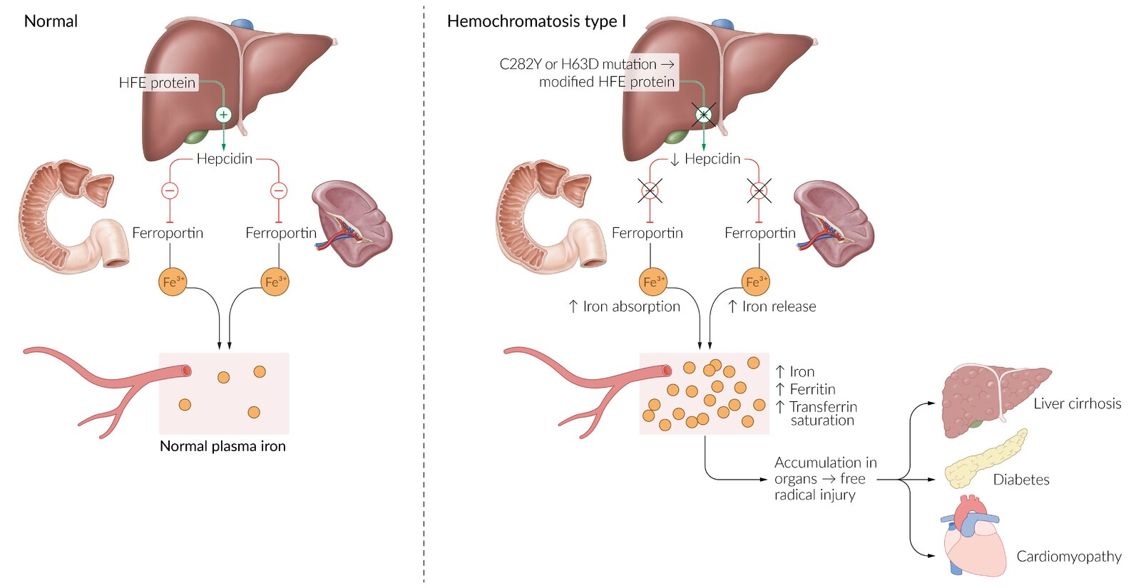
Tip
Don’t mess up with Wilson disease
- Wilson disease is accumulation of copper, and has neurologic symptoms.
Clinical features
Tip
Classic triad of cirrhosis, diabetes mellitus, skin pigmentation (“bronze diabetes”).
- Early/Constitutional: Severe fatigue, generalized lethargy, arthralgias.
- Musculoskeletal: c
- Pseudogout (CPPD) and degenerative arthropathy.
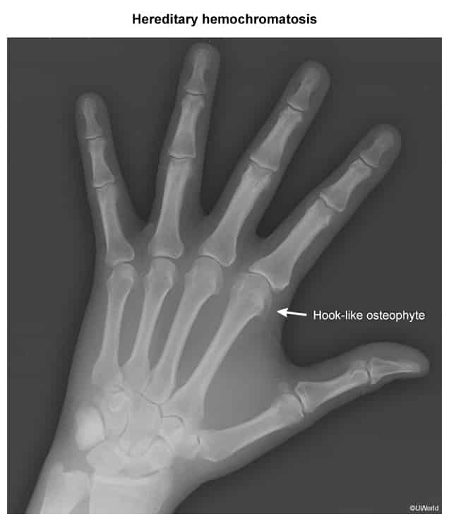
- Classically involves the 2nd and 3rd metacarpophalangeal (MCP) joints with “hook-like” osteophytes.
- Dermatologic: “Bronze” skin (hyperpigmentation due to iron deposition and stimulated melanin production).
- Gastrointestinal/Hepatic: Hepatomegaly (early), progresses to cirrhosis, jaundice, and portal HTN.
- Endocrine:
- “Bronze Diabetes”: Pancreatic islet -cell damage secondary DM.
- Secondary Hypogonadism: Iron deposition in anterior pituitary LH/FSH ED, testicular atrophy, loss of libido, amenorrhea. c
- Hypothyroidism, hypoparathyroidism (less common).
- Cardiac:
- Dilated cardiomyopathy (more common) or restrictive cardiomyopathy.
- Conduction abnormalities, sick sinus syndrome, arrhythmias (atrial fibrillation/flutter).
- Infectious Susceptibility:
- Increased risk of fulminant sepsis from siderophilic organisms:
- Vibrio vulnificus (exposure to raw oysters/warm seawater necrotizing fasciitis/bullae).
- Yersinia enterocolitica (pseudoappendicitis, bacteremia).
- Listeria monocytogenes.
- Increased risk of fulminant sepsis from siderophilic organisms:
| Feature | Hemochromatosis (Iron Overload) | Wilson Disease (Copper Overload) |
|---|---|---|
| Gene Defect | HFE gene (Autosomal Recessive) → ↑ Iron absorption. | ATP7B gene (Autosomal Recessive) → ↓ Copper excretion. |
| ”Classic” Triad / Signs | 1. Cirrhosis (↑ HCC risk) 2. Diabetes Mellitus 3. Skin Pigmentation (“Bronze Diabetes”) | 1. Liver Disease (Hepatitis, Cirrhosis) 2. Neurologic Dysfunction (Parkinsonism, tremor) 3. Kayser-Fleischer Rings (Ocular) |
| Key Labs | ↑ Ferritin ↑ Iron ↑ Transferrin Saturation | ↓ Ceruloplasmin ↑ Urinary Copper |
| Treatment | Phlebotomy Iron Chelators (Deferasirox) | Copper Chelators (Penicillamine, Trientine) Oral Zinc |
Diagnostics
- Initial/Screening:
- Iron panel: ↑ Transferrin saturation (often >45%; most sensitive initial test) and ↑ Ferritin (reflects total body iron stores).
- Confirmatory/Gold Standard:
- HFE genetic testing (C282Y/H63D mutation analysis).
- Key Labs:
- Mildly elevated AST/ALT.
- Liver biopsy
- Hemosiderin (normally golden yellow on microscopy) appears blue with the Prussian blue stain.
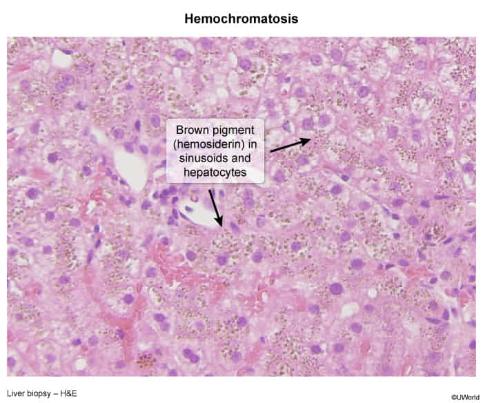
- Pattern of hereditary hemochromatosis: pronounced parenchymal siderosis (accumulation of hemosiderin within the tissue) in hepatocytes and bile duct epithelium
- Pattern of secondary iron overload: Kupffer cells (specialized macrophages) containing hemosiderin
- Hemosiderin (normally golden yellow on microscopy) appears blue with the Prussian blue stain.
Treatment
- First-line: Therapeutic phlebotomy.
- Effective and simple. Only required 2 to 4 times a year. HH patients can often perform this via standard blood donations
- Indication: Ferritin >500 ng/mL or symptomatic iron overload.
- Target: Maintain ferritin between 50–100 ng/mL and transferrin saturation <50%.
- Second-line / Alternative: Iron chelation therapy (e.g., Deferoxamine, Deferasirox).
- Indication: Patients with severe anemia, severe cardiac disease, or poor venous access preventing phlebotomy.
- Dietary Modifications:
- Avoid iron supplements, vitamin C (enhances iron absorption), and alcohol.
- Avoid raw shellfish due to high susceptibility to fatal septicemia.
- Definitive/End-stage: Liver transplantation for decompensated cirrhosis or localized HCC.