Location: Paired, oval glands located in the scrotum.
Coverings:
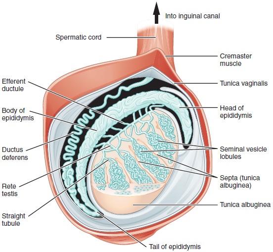
Tunica Vaginalis: Outer serous layer, derived from peritoneum. Has visceral and parietal layers. Fluid accumulation here causes a Scrotal abnormalities.
Tunica Albuginea: Inner, tough fibrous capsule that extends septa into the testis, dividing it into ~250 lobules.
Internal Structure:
Each lobule contains 1-4 seminiferous tubules (site of spermatogenesis).
Seminiferous tubules converge to form straight tubules (tubuli recti).
Straight tubules empty into the rete testis, a network of channels in the mediastinum testis.
Sperm travels from the rete testis to the epididymis via efferent ductules.
Spermatic Cord: Contains the testicular artery, pampiniform plexus, and ductus deferens.
Vasculature & Lymphatics
Arterial Supply:Testicular Artery
Arises directly from the abdominal aorta at the L2 vertebral level.
Venous Drainage:Pampiniform Plexus
A network of veins surrounding the testicular artery, important for thermoregulation (countercurrent heat exchange).
The plexus coalesces to form the testicular vein.
Right Testicular Vein drains directly into the Inferior Vena Cava (IVC).
Left Testicular Vein drains into the Left Renal Vein at a 90-degree angle, making the left side more susceptible to varicocele.
Lymphatic Drainage:
Para-aortic (lumbar) lymph nodes.
Key point:Testicular cancer metastasizes to para-aortic nodes, NOT inguinal nodes. Scrotal skin drains to superficial inguinal nodes.
Histology & Cell Types
Seminiferous Tubules: Site of spermatogenesis, containing two main cell types.
Sertoli Cells (Sustentacular Cells):
Function: Support and nourish developing sperm, form the blood-testis barrier via tight junctions, secrete inhibin B (inhibits FSH), androgen-binding protein (ABP) (concentrates testosterone), and Müllerian-inhibiting factor (MIF) in the fetus.
Hormonal Control: Stimulated by FSH.
Appearance: Columnar cells extending from the basement membrane to the lumen.
Spermatogenic (Germ) Cells: All stages of sperm development.
Spermatogonia (diploid) are located at the basal lamina.
They mature into primary spermatocytes, secondary spermatocytes, spermatids, and finally spermatozoa (sperm).
Interstitial Tissue: Connective tissue between seminiferous tubules.
Leydig Cells (Interstitial Cells):
Function: Synthesize and secrete testosterone in response to LH.
Appearance: Eosinophilic cytoplasm, often found in clusters. May contain rod-shaped cytoplasmic crystals of Reinke.
Hormonal Control: Stimulated by LH.
Clinical Correlations
Testicular Torsion: Twisting of the spermatic cord, leading to ischemia. A surgical emergency characterized by severe pain and an absent cremasteric reflex.
Varicocele: Dilation of the pampiniform plexus (“bag of worms” feel). Much more common on the left side due to the left testicular vein draining into the left renal vein.
Hydrocele: Collection of peritoneal fluid within the tunica vaginalis. Transilluminates on physical exam.
Cryptorchidism: Undescended testis, which can lead to infertility and an increased risk of testicular cancer.