Placental abruption
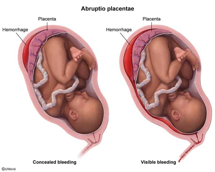
The partial or complete separation of the placenta from the uterus prior to delivery; subsequent hemorrhage occurs from both maternal and fetal vessels.
Epidemiology
- Occurs most often in the third trimester
Etiology
- Pathophysiology: Premature detachment of placenta from uterine decidua before fetal delivery rupture of maternal spiral arteries decidual hematoma.
- Major Risk Factors:
- Maternal HTN / Preeclampsia / Chronic HTN (strongest overall predictor). c
- Abdominal trauma (e.g., MVA, domestic violence) / Rapid uterine decompression (e.g., ROM in polyhydramnios, delivery of twin A).
- Cocaine or amphetamine use (vasospasm ischemia).
- Tobacco smoking.
- Prior history of placental abruption.
- Advanced maternal age (AMA) and high parity.
- Uterine anomalies, leiomyomas (especially submucosal).
Clinical features
- Classic Presentation: Painful 3rd-trimester vaginal bleeding + hypertonic, tender uterus (“woody” abdomen) + high-frequency contractions (tachysystole).
- Concealed Abruption (~20%): Bleeding is entirely retroplacental; no visible vaginal bleeding, but severe abdominal pain, uterine hypertonus, and maternal hemodynamic instability are present. c
- Fetal Tracing: Fetal distress (loss of variability, recurrent late decelerations, bradycardia, sinusoidal pattern).
- PE: Uterine tenderness on palpation, elevated resting uterine tone, maternal hypotension/tachycardia.
Management
- Stabilization (Immediate):
- 2 large-bore IVs, aggressive IVF, crossmatched PRBCs/FFP/cryo, O2, left lateral tilt.
- Administer Rh(D) immune globulin if Rh-negative.
- Emergency C-Section:
- Indicated for maternal hemodynamic instability, DIC, or non-reassuring FHR.
- Vaginal Delivery:
- Indicated if mother and fetus are stable with term/rapidly progressing labor.
- Expectant Management:
- Preterm (< 34 weeks), mild/stable abruption, reassuring FHR; give antenatal steroids (betamethasone).
Placenta previa
Presence of the placenta in the lower uterine segment, which can lead to partial or full obstruction of the internal os; high risk of hemorrhage (rupture of placental vessels) and birth complications
Risk factors
- Maternal age > 35 years, multiparity, short intervals between pregnancies
- Previous curettage or cesarean delivery
- Previous placenta previa, previous/recurrent abortions
Classification
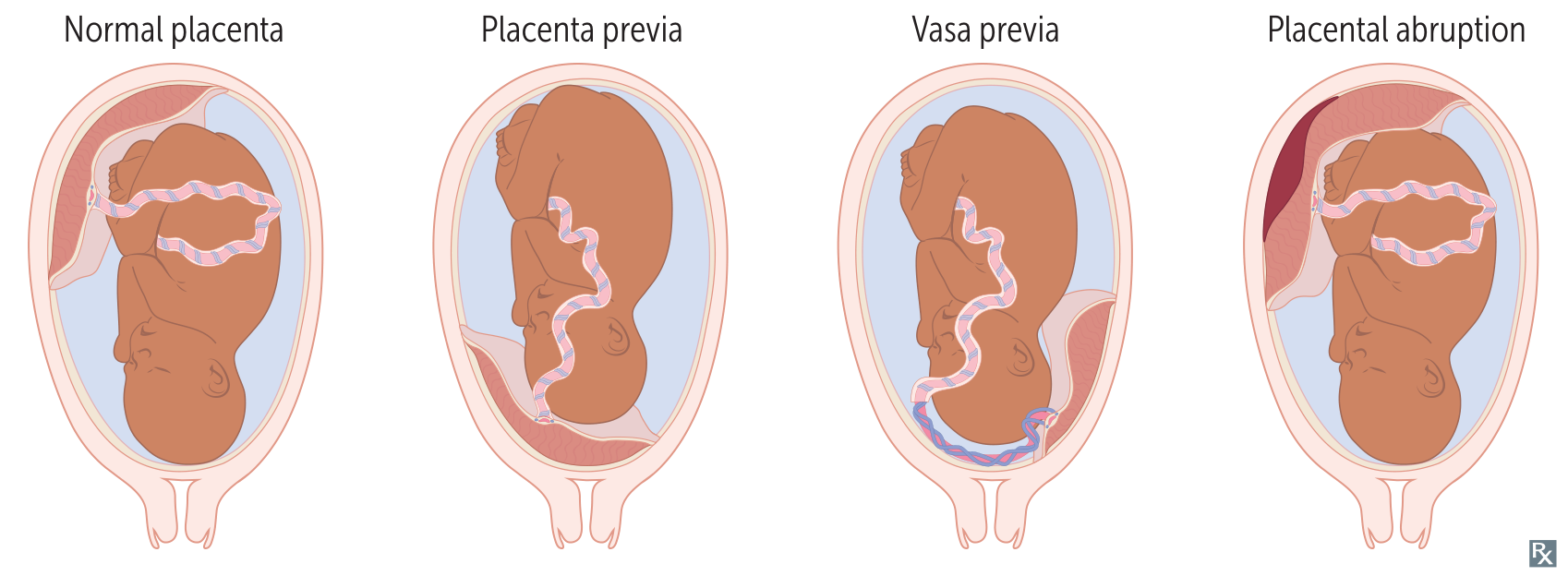
- Placenta previa: placenta either partially or completely covers the internal os
- Previously, this category included marginal previa (placenta reaching the internal os), partial previa (placenta partially covering the internal os), and complete previa (placenta completely covering the internal os); these terms have been excluded from the new classification.
- Low-lying placenta: lower edge of the placenta lies less than 2 cm from the internal cervical os
Clinical features
- Sudden, painless, bright red vaginal bleeding
- Usually occurs during the 3rd trimester (before rupture of the membranes)
- Initial bleeding episodes are often self-limited and recur during the onset of labor
- Soft, nontender uterus
- Usually no fetal distress
Treatment
- Asymptomatic/Stable: Expectant management with pelvic rest (no intercourse, no digital exams), and serial ultrasounds. Corticosteroids (e.g., betamethasone) are given for fetal lung maturity if <34 weeks.
- Active Bleeding:
- Unstable mother or fetus: Immediate C-section regardless of gestational age.
- Stable mother and fetus (>36 weeks): Scheduled C-section is the standard mode of delivery.
- Never deliver vaginally if there’s a complete previa.
- IV access with large-bore cannulas, blood type and crossmatch are essential due to hemorrhage risk.
Vasa previa
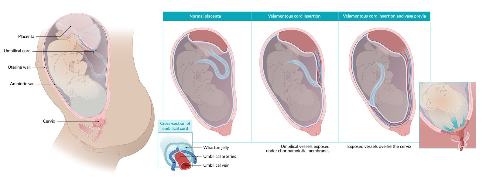
- Definition & Pathophysiology: Unprotected fetal blood vessels traverse membranes over the internal cervical os without Wharton jelly support (high risk of rupture/compression).
- Risk Factors: Velamentous cord insertion, succenturiate/accessory lobe, low-lying placenta, IVF, twins.
- Clinical Presentation (Classic Triad):
- Painless vaginal bleeding
- Triggered by rupture of membranes (ROM)
- Fetal distress (fetal bradycardia, sinusoidal FHR pattern) w/ stable maternal vitals.
- Diagnosis:
- Prenatal/Gold Standard: TVUS w/ Color Doppler (shows fetal vessels directly overlying/within 2 cm of os).
- Intrapartum: Apt test or Wright stain (detects fetal HbF vs maternal HbA; usually skipped for immediate delivery).
- Differential Diagnosis: c
- Placenta Previa: Painless bleeding before ROM; maternal origin (maternal shock); normal initial FHR.
- Placental Abruption: Painful bleeding + uterine tenderness/hypertonus.
- Uterine Rupture: Severe abdominal pain, loss of fetal station, maternal collapse, prior C-section scar.
- Management:
- Prenatal (Diagnosed early): Hospitalize at 30–34 wks -> Antenatal corticosteroids -> Elective C-section at 34–35 wks.
- Emergency (Active bleeding / ROM / Fetal distress): Immediate Emergency C-section + prepare O-neg PRBCs for neonatal transfusion.