| Disorder | Key Gene / Locus | Primary Skin Finding | Primary CNS Finding | ”Can’t Miss” Association |
|---|---|---|---|---|
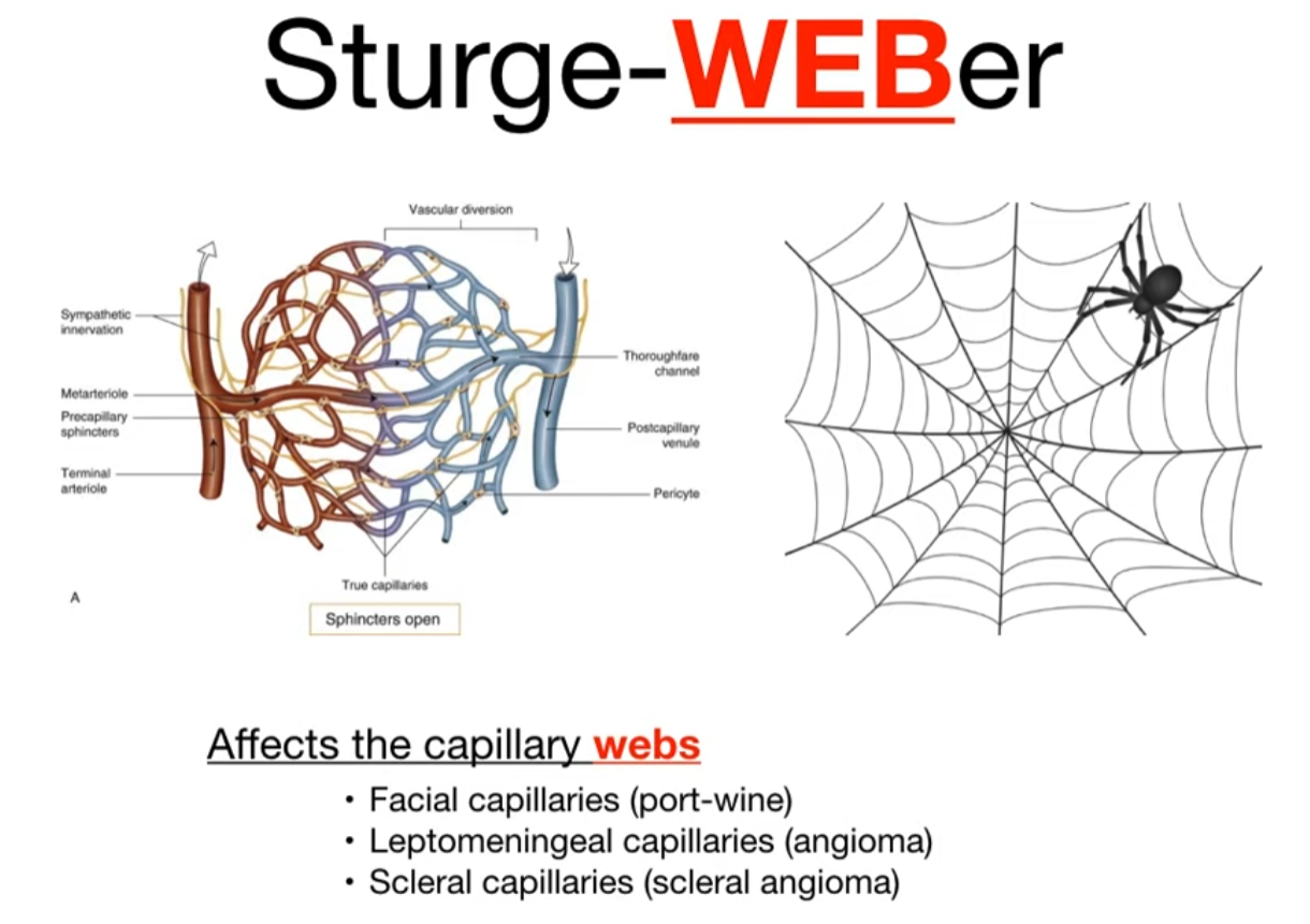
| Sturge-Weber | Sporadic (GNAQ) | Port-Wine Stain (V1/V2) | Leptomeningeal Angioma (“Tram-track” calcifications) | Glaucoma |
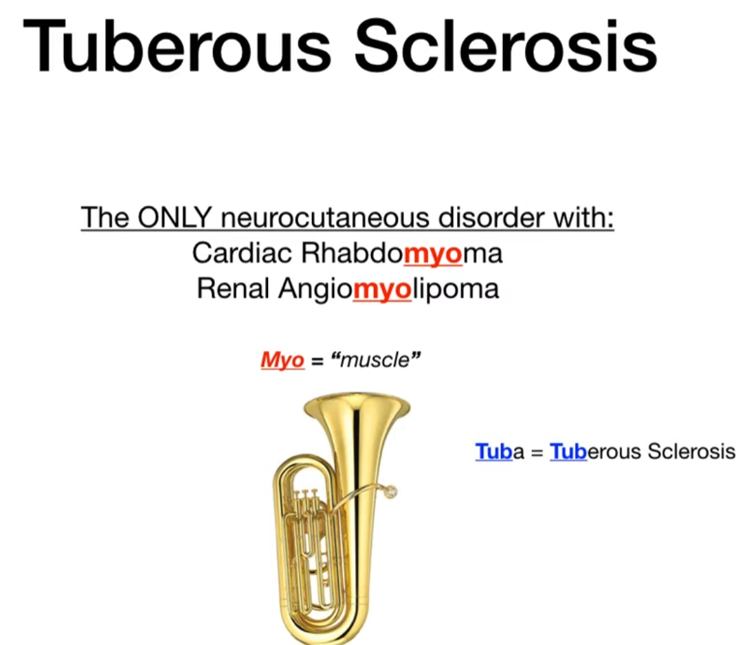
| Tuberous Sclerosis | TSC1/TSC2 | Ash-leaf spots, Angiofibromas | Cortical Tubers, Subependymal giant cell astrocytomas (SEGAs) | Cardiac Rhabdomyoma, Renal Angiomyolipoma |
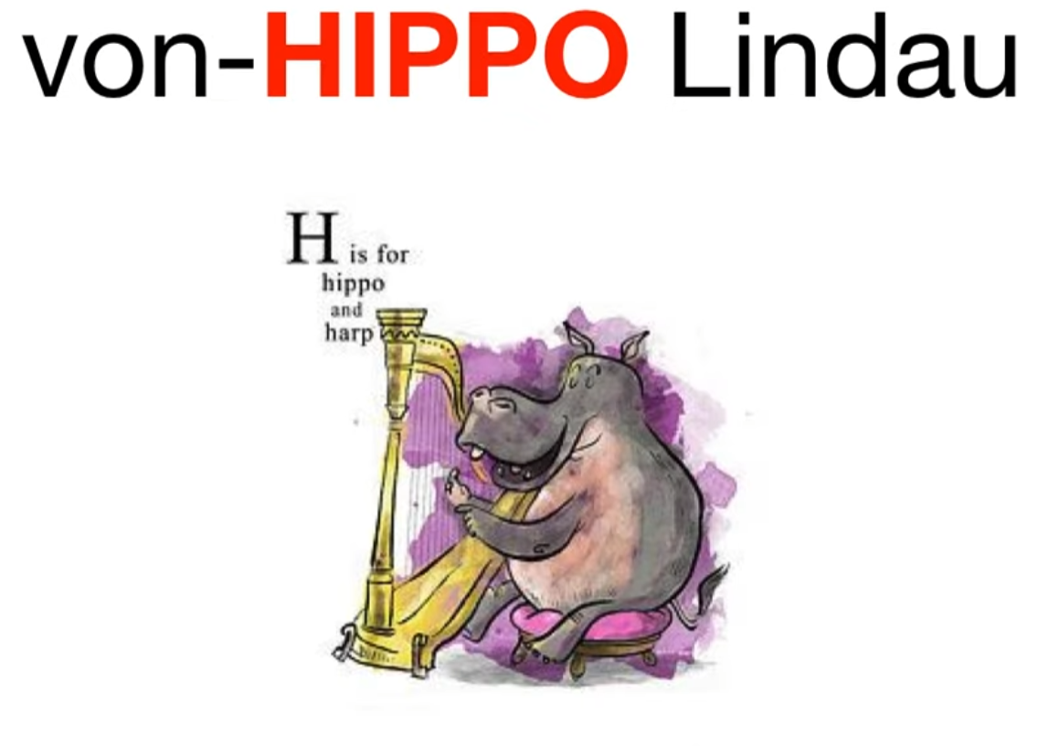
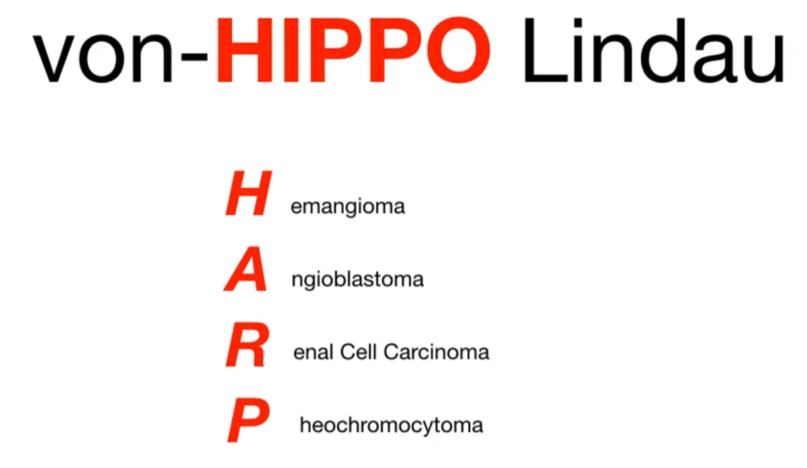
| von Hippel-Lindau | VHL (Chr 3) | (Minimal skin findings) | Hemangioblastomas (retina, cerebellum) | Renal Cell Carcinoma, Pheochromocytoma |
| NF1 | NF1 (Chr 17) | Café-au-lait spots, Cutaneous neurofibromas Axillary freckling | Optic Glioma | Lisch nodules (iris hamartomas) |
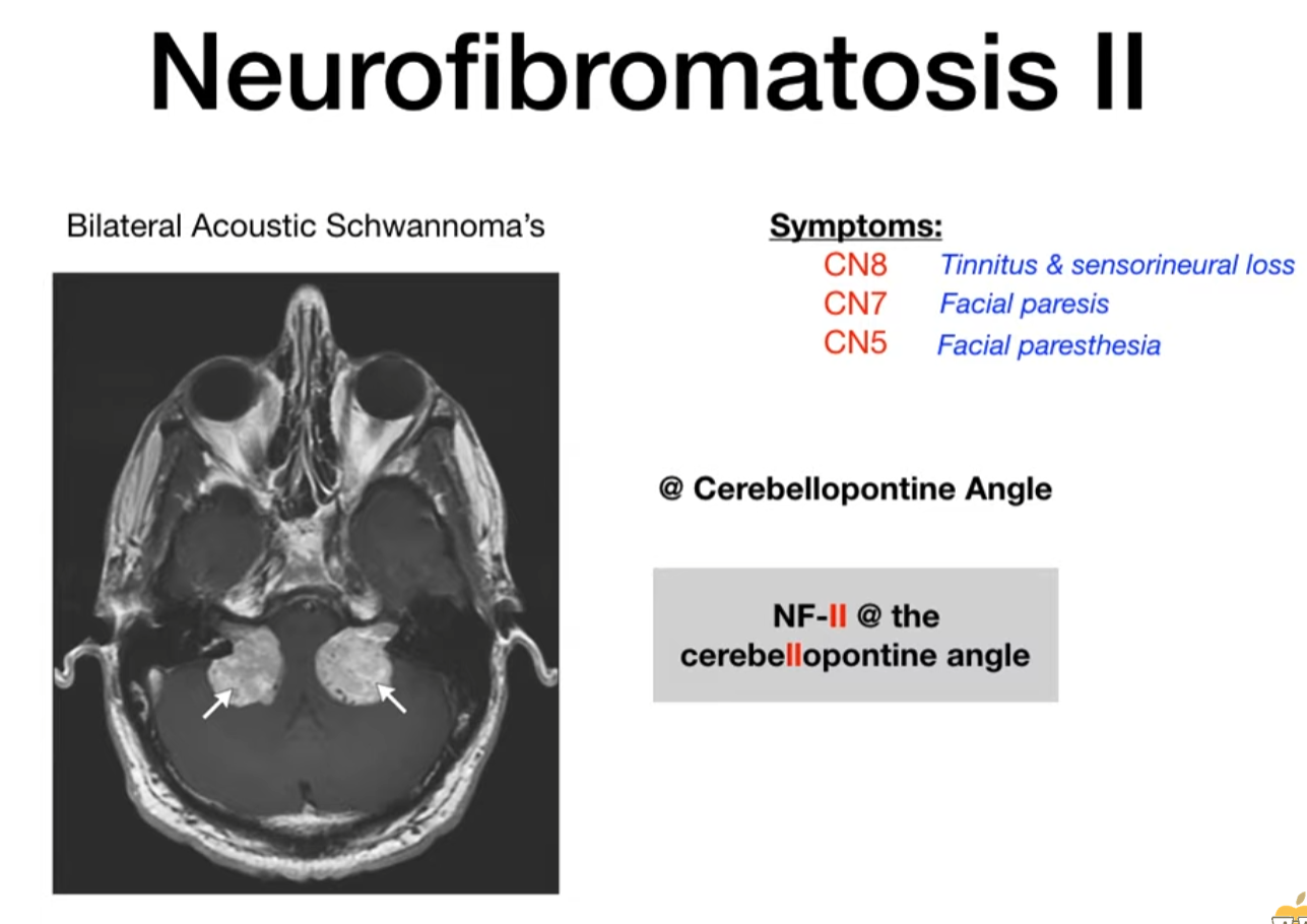
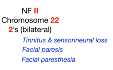
| NF2 | NF2 (Chr 22) | (Fewer/milder skin findings) | Bilateral Vestibular Schwannomas | Multiple meningiomas, Ependymomas |
Sturge-Weber syndrome
- Pathophysiology/Etiology
- A rare, sporadic neurocutaneous disorder also known as encephalotrigeminal angiomatosis.
- Caused by a somatic mosaic mutation in the GNAQ gene during early development. This is not an inherited condition.
- The mutation leads to abnormal development and persistence of embryonal blood vessels, resulting in capillary-venous malformations in the skin, brain, and eye.
- Clinical Presentation
- Classic Triad: 1) Facial capillary malformation (port-wine birthmark), 2) Leptomeningeal angioma, and 3) Ocular abnormalities (like glaucoma).
- Skin: A congenital, unilateral port-wine birthmark (nevus flammeus) is the hallmark, typically in the V1 (ophthalmic) distribution of the trigeminal nerve. The likelihood of SWS increases if the V1 distribution is involved.
- Neurologic: Seizures are the most common neurologic feature, often focal and starting in the first year of life. Other findings include developmental delay, intellectual disability, hemiparesis, and headaches.
- Ocular: Glaucoma is a major concern (occurring in 30-70% of patients) and can lead to optic nerve damage and blindness. Other findings include choroidal hemangiomas and buphthalmos (enlargement of the eyeball). c
Tuberous sclerosis
- Inheritance: Autosomal Dominant (AD); ~66% cases due to de novo mutations.
- Pathophysiology: Loss-of-function mutation in tumor suppressor genes TSC1 (Hamartin, Chr 9) or TSC2 (Tuberin, Chr 16) constitutive activation of the mTOR pathway widespread benign hamartomas in multiple organ systems.
- Clinical features
- Cutaneous Findings:
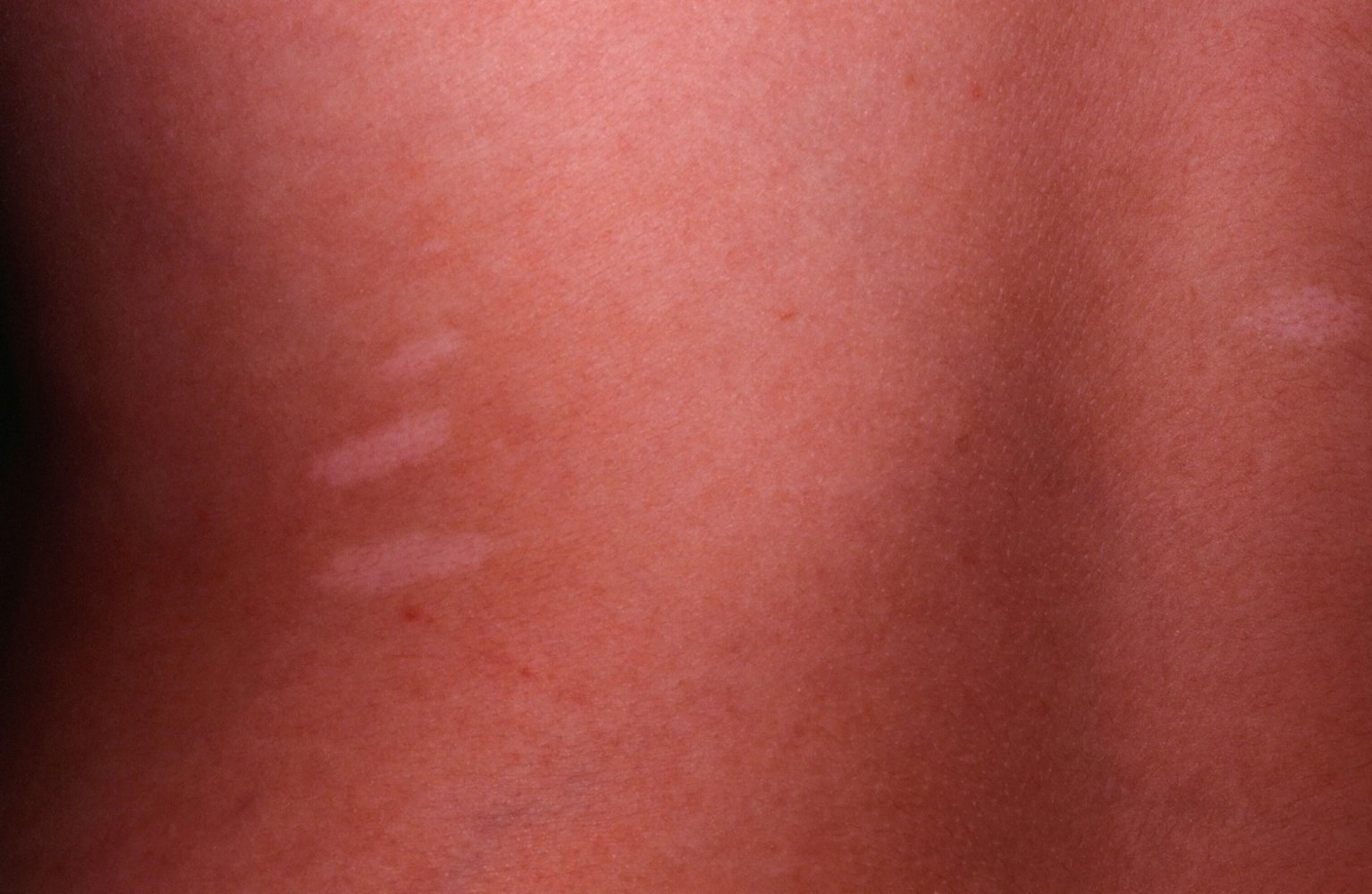
- Ash-leaf spots: Hypopigmented macules (earliest sign, best seen via Wood lamp).
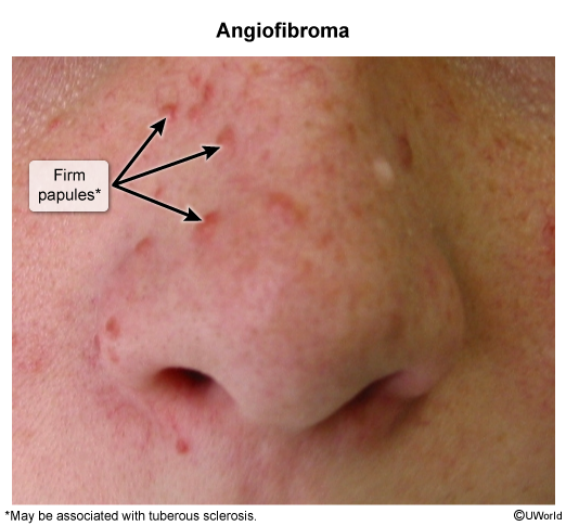
- Angiofibromas (Adenoma sebaceum): Malar erythema/papules appearing in early childhood; spares nasolabial folds.
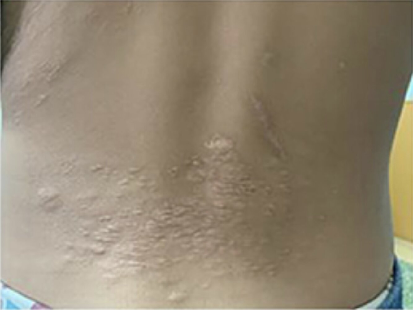
- Shagreen patch: Leathery, textured plaque usually in the lumbosacral area.
- Periungual/Ungual fibromas (Koenen tumors): Flesh-colored nodules under/around nail beds (develop in adolescence/adulthood).
- Ash-leaf spots: Hypopigmented macules (earliest sign, best seen via Wood lamp).
- Neurological Manifestations:
- Seizures: Infantile spasms (hypsarrhythmia on EEG) in infancy; generalized or focal seizures later. c
- Subependymal Giant Cell Astrocytoma (SEGA): Can cause obstructive hydrocephalus (headache, vomiting, papilledema). c
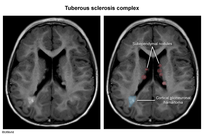
- Subependymal nodules (SEN) and Cortical tubers: Cause epilepsy, Intellectual Disability (ID), and Autism Spectrum Disorder (ASD).
- Cardiovascular:
- Cardiac Rhabdomyoma: Often detected prenatally or in infancy; typically spontaneously regresses; may cause arrhythmias or outflow tract obstruction.
- Renal:
- Renal Angiomyolipoma (AML): Often bilateral and multiple; high risk of spontaneous retroperitoneal hemorrhage if >4\text{ cm}.
- Renal cysts and increased risk of early-onset Renal Cell Carcinoma (RCC).
- Pulmonary:
- Lymphangioleiomyomatosis (LAM): Almost exclusively in post-pubertal females; manifests as dyspnea, recurrent spontaneous pneumothorax, and cystic lung destruction.
- Ocular:
- Retinal hamartomas (astrocytic hamartoma): Asymptomatic, “mulberry-like” lesions on fundoscopy.
- Cutaneous Findings:
von Hippel-Lindau syndrome
- Patho/Etiology
- Autosomal dominant mutation of the VHL tumor suppressor gene on chromosome 3p.
- Loss of VHL protein function leads to impaired ubiquitination and elimination of hypoxia-inducible factor 1α (HIF-1α).
- Constitutive upregulation of HIF-1α promotes the transcription of genes for angiogenesis (e.g., VEGF, PDGF) and erythropoietin, leading to the formation of highly vascular tumors.
- Clinical Presentation
- Multisystem disorder characterized by the development of numerous benign and malignant tumors and cysts.
- Onset typically in young adulthood (average age 26).
- Common manifestations include:
- Hemangioblastomas: Most common tumors. Found in the CNS (especially cerebellum and spine) and retina. Cerebellar lesions can cause ataxia, and headaches. c2
- Renal Cell Carcinoma (RCC): Specifically clear cell subtype, often bilateral and multifocal. Occurs in up to 70% of patients and is the leading cause of mortality. c
- Not renal hemangioma
- Pheochromocytomas: Can be bilateral and cause episodic or sustained hypertension. c
- Pancreatic Lesions: Include simple cysts, serous cystadenomas, and neuroendocrine tumors (pNETs).
- Endolymphatic Sac Tumors: Located in the inner ear, can cause hearing loss.
- Epididymal and Broad Ligament Cystadenomas.
- See Hereditary cancer syndromes
Neurofibromatosis
- Etiology
- Neurofibromatosis type 1 and type 2: autosomal dominant inheritance or spontaneous mutation t
Neurofibromatosis type I (Peripheral Neurofibromatosis)
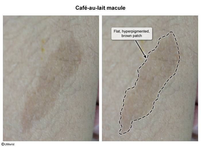
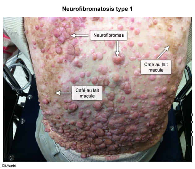
- Café-au-lait spots
Mnemonic
If you drink too much coffee, you are gonna go number one.
- Café-au-lait spots: ≥6 macules (>5 mm prepubertal, >15 mm postpubertal).
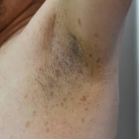
- Axillary/inguinal freckling (Crowe sign). c
- Neurofibromas: ≥2 cutaneous/subcutaneous neurofibromas OR ≥1 plexiform neurofibroma (pathognomonic, risk of malignant transformation).
- Cutaneous neurofibromas are benign nerve sheath tumors composed of diverse cells and are often found within the dermis. The dermis is composed of collagen, elastic fibers, and ground substance and contains extracellular components including nerves, vasculature, hair follicles, and glands. Neurofibromas are composed of a mixture of cells normally found in peripheral nerves, including neoplastic Schwann cells, as well as non-neoplastic fibroblasts, perineural cells, and mast cells.
- Cutaneous neurofibromas are benign nerve sheath tumors composed of diverse cells and are often found within the dermis. The dermis is composed of collagen, elastic fibers, and ground substance and contains extracellular components including nerves, vasculature, hair follicles, and glands. Neurofibromas are composed of a mixture of cells normally found in peripheral nerves, including neoplastic Schwann cells, as well as non-neoplastic fibroblasts, perineural cells, and mast cells.
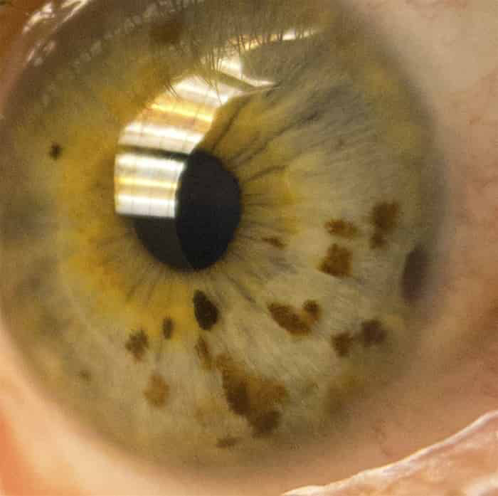
- Lisch nodules: Pigmented iris hamartomas (asymptomatic, seen on slit-lamp).
- Optic pathway glioma: Vision loss, proptosis, precocious puberty (if hypothalamic involvement). c2
- Osseous lesions: Sphenoid wing dysplasia, pseudoarthrosis (anterolateral tibial bowing), severe scoliosis.
- Other: Cognitive impairment/learning disabilities, HTN (secondary to renal artery stenosis or pheochromocytoma).
Neurofibromatosis type II (Central Neurofibromatosis)
- Bilateral vestibular schwannomas (acoustic neuromas): Tinnitus, sensorineural hearing loss (SNHL), vertigo, ataxia. c
- Multiple intracranial/spinal tumors: Meningiomas, ependymomas, schwannomas.
- Ocular: Juvenile posterior subcapsular cataracts, retinal hamartomas (leads to early vision impairment).
- Skin lesions: Few cutaneous schwannomas, plaque-like lesions (lacks classic café-au-lait/freckling density of NF1).