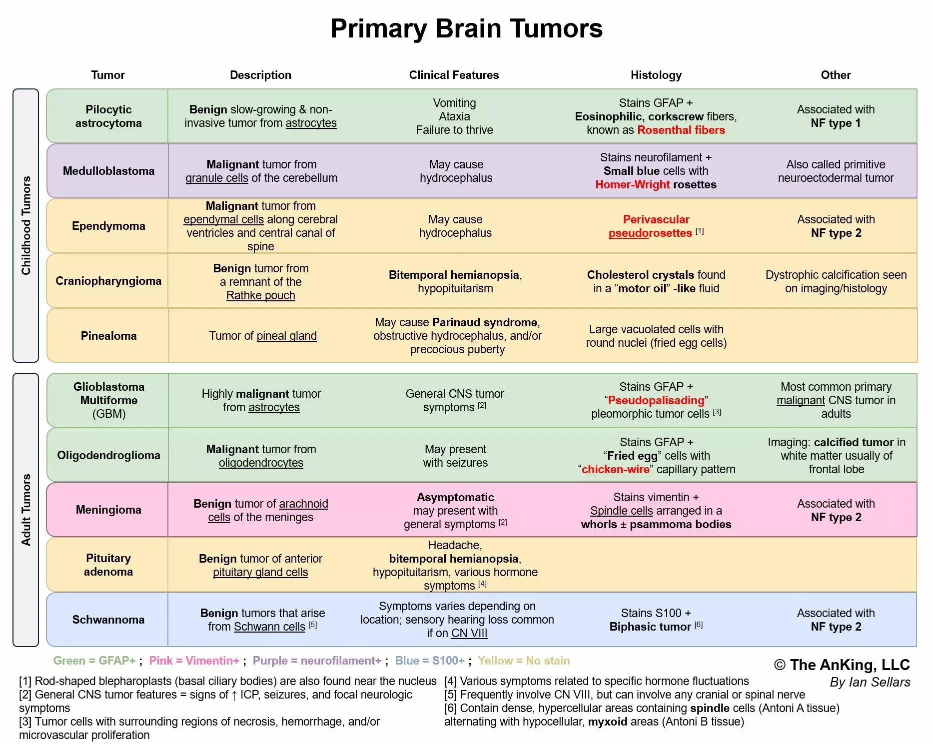
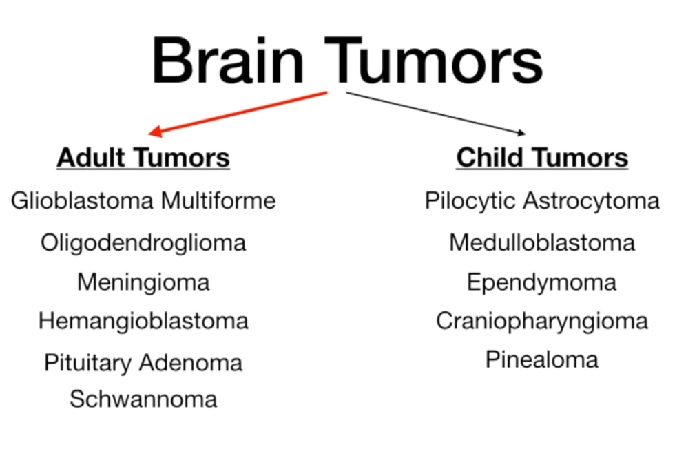
- Adult Brain Tumors (Supratentorial)
- Glioblastoma: Most common primary malignant. Butterfly glioma (crosses corpus callosum), pseudopalisading necrosis.
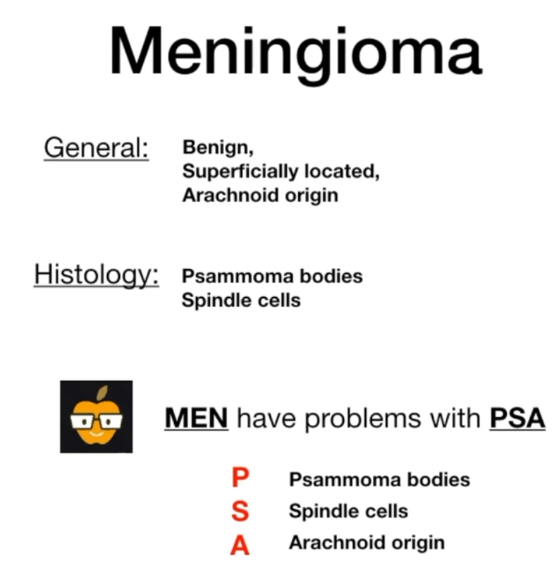
- Meningioma: 2nd most common. Benign, dural-based. Psammoma bodies, whorled pattern.
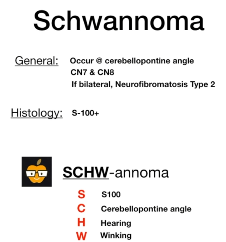
- Schwannoma: Cerebellopontine angle (CPA), affects CN VIII. S-100 positive. Bilateral = NF2.
- Oligodendroglioma: “Fried-egg” cells, “chicken-wire” capillaries, often calcified.
- Pediatric Brain Tumors (Infratentorial)
- Pilocytic Astrocytoma: Most common, benign. Cerebellum. Rosenthal fibers, cystic lesion with mural nodule.
- Medulloblastoma: Most common malignant. Cerebellar vermis. Homer-Wright rosettes, small round blue cells, risk of drop metastases.
- Ependymoma: 4th ventricle → hydrocephalus. Perivascular pseudorosettes.
- Craniopharyngioma: From Rathke’s pouch. Suprasellar, calcified, contains cholesterol crystals. Causes bitemporal hemianopsia.
Site
- Pediatric Tumors → Infratentorial (Posterior Fossa)
- Reason: The cerebellum and brainstem undergo significant development and cell proliferation late in gestation and in the first few years of life. A higher rate of cell division and migration increases the chance of oncogenic mutations.
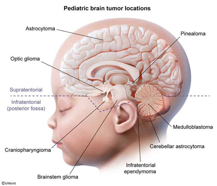
- Reason: The cerebellum and brainstem undergo significant development and cell proliferation late in gestation and in the first few years of life. A higher rate of cell division and migration increases the chance of oncogenic mutations.
- Adult Tumors → Supratentorial (Cerebral Hemispheres)
- Reason: By adulthood, the rapid developmental proliferation has ceased. Tumors now arise from the supportive (glial) and covering (meningeal) cells that are maintained throughout life.
Tip
Most tumors are glial origin, except for medulloblastoma (neural origin).
Adult tumors
Glioblastoma multiforme
- Pathophysiology
- Most common primary malignant brain tumor in adults.
- World Health Organization (WHO) Grade IV astrocytoma.
- Arises from astrocytes. Highly aggressive and infiltrative.
- Commonly found in cerebral hemispheres. Can cross the corpus callosum, creating a “butterfly glioma”.
- Clinical Features
- Pt Profile: Typically > 50 years old.
- Rapidly progressive neurologic symptoms over weeks to months.
- Symptoms depend on tumor location: focal deficits (e.g., weakness, aphasia), cognitive changes.
- General Sx of ↑ Intracranial Pressure (ICP): headache (often worse in the morning), nausea/vomiting, papilledema. c
- Worse in the morning: due to supine position and CO₂ retention during sleep
- Seizures are a common presenting symptom.
- Diagnostics
- MRI with contrast is the imaging modality of choice.
- Classic finding: Irregular, thick-walled, ring-enhancing lesion with central necrosis and significant surrounding vasogenic edema.
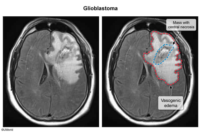
- Cause: Disruption of the Blood-Brain Barrier (BBB). t
- Mechanism: Glioblastoma cells secrete VEGF → promotes formation of abnormal, leaky blood vessels → plasma fluid extravasates into brain parenchyma.
- Result: Accumulation of protein-rich fluid in the extracellular space, primarily in white matter tracts, leading to mass effect and ↑ ICP.
- Location: Typically supratentorial (cerebral hemispheres).
- Pathology
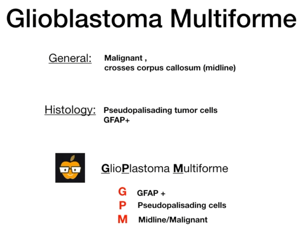
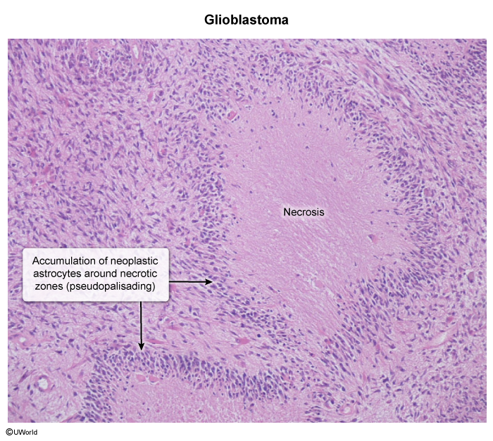
- Histologic hallmark: Pseudopalisading necrosis, where pleomorphic tumor cells line up along a central area of necrosis.
- Marked vascular/endothelial proliferation.
- Tumor cells stain positive for GFAP (Glial Fibrillary Acidic Protein).
Tip
- True Palisading (in Schwannoma): Cells actively arrange themselves in an organized way (e.g., around vessels or fibrillary cores).
- Pseudopalisading (in GBM): Cells are reactively compacted around an area of necrosis, primarily due to the tumor outgrowing its blood supply and cells migrating away from the dead center.
Oligodendroglioma
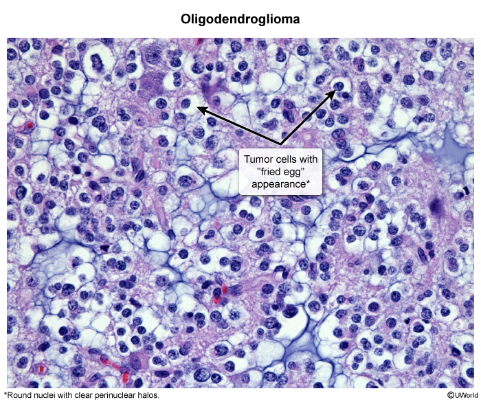
"O" is like an egg

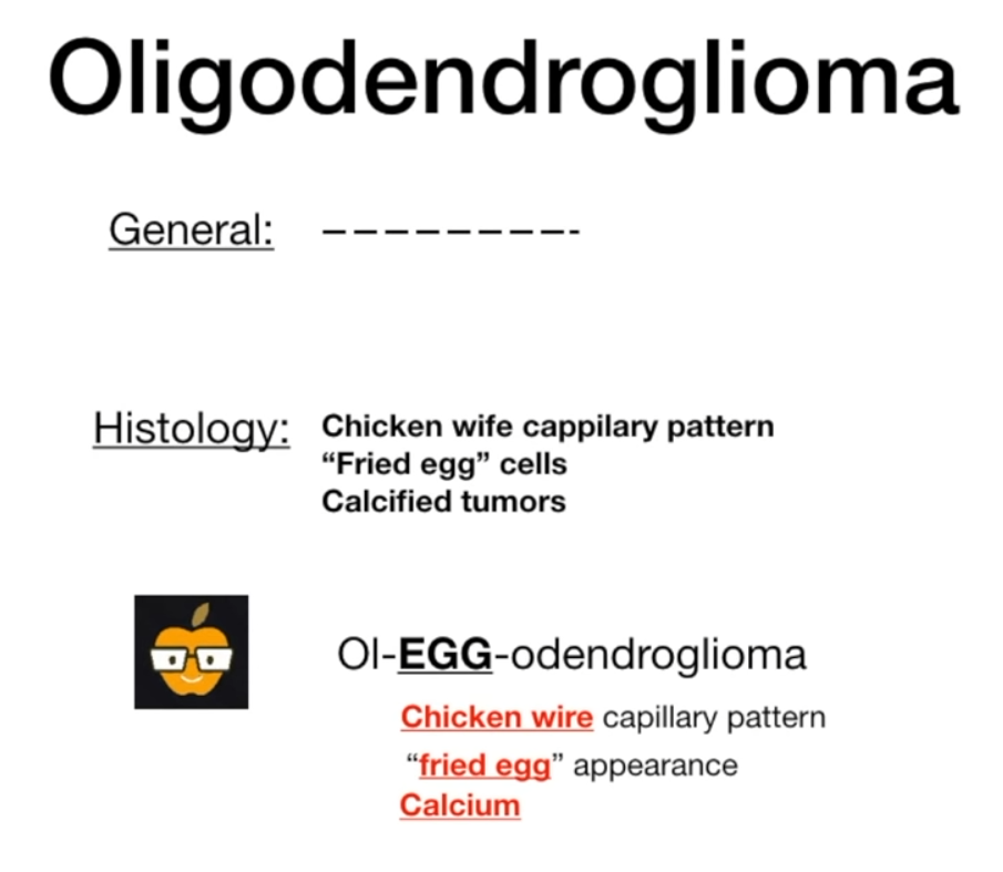 Eat egg will increase your calcium.
Meningioma
Schwannoma
Childhood tumors
Pilocytic astrocytoma
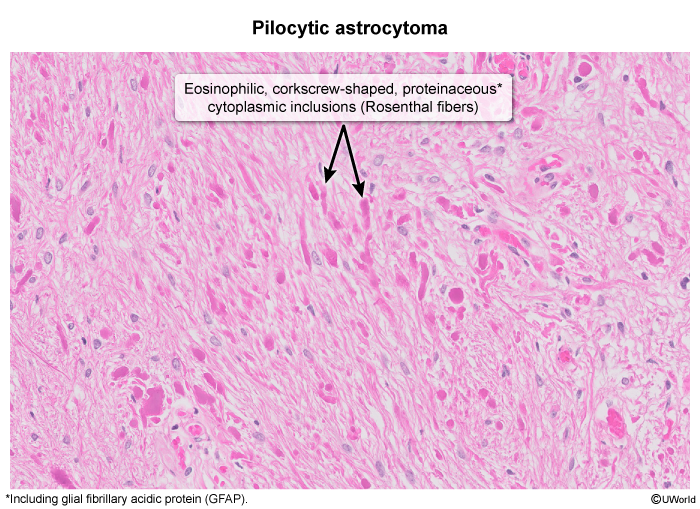 Eosinophilic fibers with corkscrew appearance (Rosenthal fibers)
Mnemonic
astROSEcytoma
Clinical Features
- Infratentorial / Cerebellar features (most common):
- Appendicular ataxia (ipsilateral dysmetria, dysdiadochokinesia, intention tremor). c
- Gait instability, wide-based gait, falls.
- Signs of ↑ ICP (secondary to mass effect or obstructive hydrocephalus via 4th ventricle compression):
- Early morning headaches, nausea, and projectile vomiting.
- Papilledema, abducens nerve (CN VI) palsy (false localizing sign).
- Lethargy, altered mental status.
- Optic Pathway Involvement (optic glioma):
- Progressive, painless vision loss, visual field cuts, proptosis, strabismus.
- Associated conditions: neurofibromatosis type I
- Supratentorial features (less common):
- Focal neurologic deficits, new-onset seizures.
Medulloblastoma
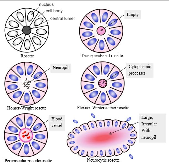
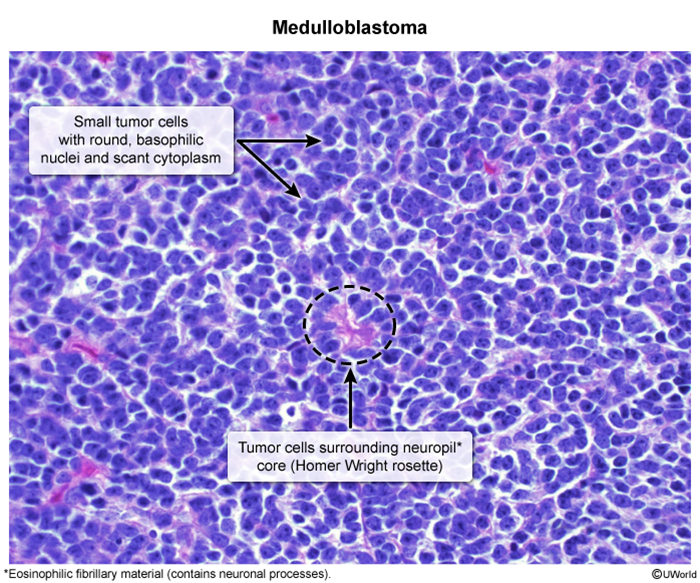
- highly malignant tumor
- Biopsy: anaplastic small round blue cells that surround a central neuropil (Homer-Wright rosettes)
Mnemonic
- Malignant and small blue cells -> Med-Blue-oblastoma
- Also remember the “Homer Wright” as “home right”, because the tumor is so malignant that there is nothing doctors can do except for sending patients right to home without hesitation.
Ependymoma
- Description: a tumor that arises from ependymal cells of the ventricular system
- Associated conditions: NF2
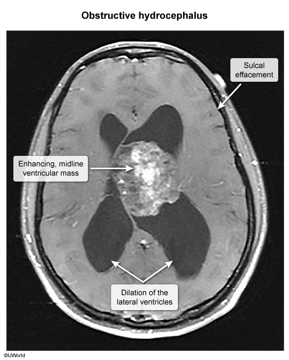
- Clinical features: the 4th ventricle is the most common location in children → noncommunicating hydrocephalus → features of increased intracranial pressure (e.g., papilledema, headache)
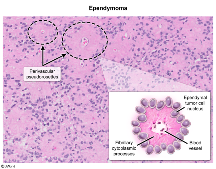
- Biopsy
- Perivascular pseudorosettes
- Rod-shaped bodies (blepharoplasts) near the nucleus
Mnemonic
“Momma’s pending gift is pseudo roses” (to make up for being a crap kid)…
Craniopharyngioma
- a benign dysontogenetic tumor arising from a remnant of the Rathke pouch (ectodermal derivative)
- Rathke pouch eventually develops into the anterior pituitary.
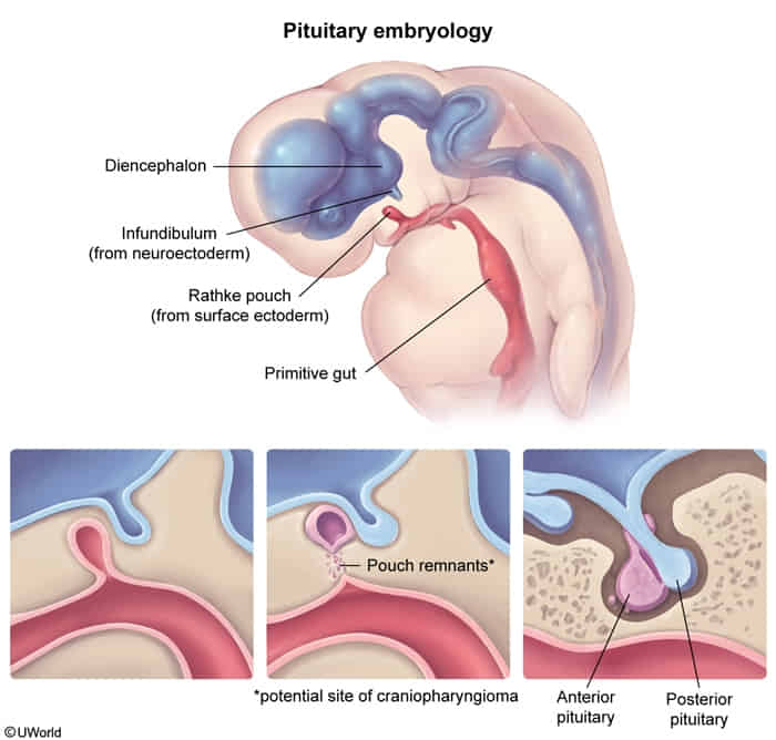
- Rathke pouch eventually develops into the anterior pituitary.
- Biopsy: cholesterol crystals found in a motor oil-like fluid on gross examination
Mnemonic
For Craniopharyngioma what i came up with was “remnants of WRATH” and i imagine a story like thats a gang or a motorcylcist group whose logo is a cranium.
Pineal gland tumors
- Clinical Presentation:
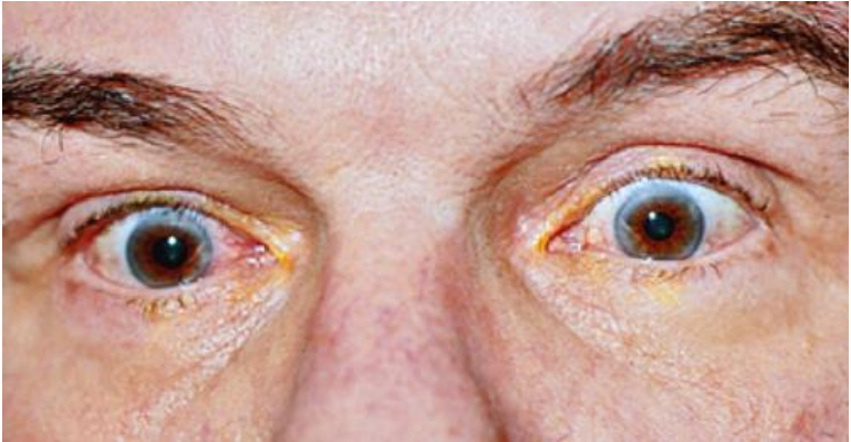
- Parinaud Syndrome (compression of pretectal area & superior colliculi): c
- Impaired upward gaze (vertical gaze palsy).
- Bilateral pupillary light-near dissociation (react to accommodation, not to light).
- Convergence-retraction nystagmus and lid retraction (Collier sign).
- Obstructive Hydrocephalus (compression of Aqueduct of Sylvius):
- Signs of ↑ ICP: Morning HA, projectile vomiting, papilledema, lethargy, CN VI palsy.
- Endocrine Effects:
- Precocious puberty in young boys (ectopic β-hCG secretion → stimulates Leydig cells → ↑ testosterone).
- Diabetes insipidus (if extending to pituitary/hypothalamus).
- Ataxia: Compression of superior cerebellar peduncles.
- Parinaud Syndrome (compression of pretectal area & superior colliculi): c
Mnemonic
- Pineal gland tumors -> Parinaud syndrome
- Consider Parinaud’s syndrome when you find someone’s peerin’ odd.
Spinal tumors
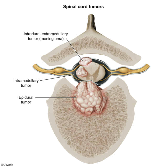
- Intramedullary space: space inside the spinal cord itself. Examples include primary spinal tumors (eg, ependymoma, astrocytoma).
- Intradural extramedullary space: space within the meninges (ie, inside the dura but outside the spinal cord). Examples include nerve sheath tumors (eg, schwannoma, neurofibroma) and tumors that originate from the meninges (eg, meningiomas).
- Extradural space: everything outside the dura, which includes the epidural space and the osseous vertebral body. Examples include vertebral metastases.
Brain metastases
- Etiology & Epidemiology:
- Incidence: Most common adult intracranial tumor (Lung > Breast > Melanoma > Colon > RCC).
- Multiplicity: Multiple = Lung, Melanoma; Solitary = Breast, Colon, RCC.
- Hemorrhagic metastases: Melanoma, RCC, Choriocarcinoma, Thyroid (MR. CT).
- Location: Grey-white matter junction (vessel narrowing traps tumor emboli).
- Clinical Presentation:
- ↑ Intracranial Pressure (ICP): Morning headache (worse with Valsalva/bending), nausea/vomiting, papilledema.
- Neurologic deficits: Focal neurologic deficits (FND), cognitive decline, or new-onset seizure in adults >40 yo. c
- Diagnosis:
- Gold standard / Best initial: Brain MRI w/ IV contrast well-circumscribed, ring-enhancing lesions located at the grey-white matter junction with severe vasogenic edema (T2/FLAIR hyperintensity). c
- Emergency / Initial: Non-contrast head CT (excludes acute bleed, mass effect, or midline shift).
- Unknown primary workup: CT Chest/Abdomen/Pelvis w/ contrast, mammography, skin exam.
- Key Differentials:
- Glioblastoma (GBM): Infiltrative, thick/irregular margins, crosses corpus callosum (“butterfly glioma”).
- Brain Abscess: Restricted diffusion on DWI MRI (hyperintense core), fever, contiguous infection source.
- Primary CNS Lymphoma: Periventricular, CSF EBV DNA (+) in HIV ().
- Management:
- Acute Vasogenic Edema / ↑ ICP: IV Dexamethasone immediately; add Mannitol / 30° head elevation if herniation threatens.
- Seizures: Levetiracetam for active seizures (no routine prophylaxis if non-seizing).
- Single accessible lesion (good functional status / KPS > 70): Surgical resection + Stereotactic Radiosurgery (SRS) to cavity (or SRS alone if deep/unresectable).
- Oligometastases (2–4 lesions): Stereotactic Radiosurgery (SRS).
- Multiple / Diffuse lesions (>4–5) or poor performance status: Whole-Brain Radiation Therapy (WBRT) targeted systemic therapy.
- Complications:
- Uncal herniation: Compression of CN III (ipsilateral fixed/dilated pupil) + contralateral hemiparesis.
- Intratumoral hemorrhage: Sudden apoplexy (highest in melanoma, RCC).
- Obstructive hydrocephalus: Compression of 4th ventricle by posterior fossa lesions.