Epidemiology & Risk Factors
- Prevalence: ~2–4% of pediatric population.
- Normal variant: Intermittent strabismus is normal in infants < 4 months of age (due to immature ocular motor control). Persistence ≥ 4 months is pathologic.
- Risk Factors:
- Prematurity / Low birth weight (LBW).
- Positive family Hx.
- Neurologic conditions: Cerebral palsy (CP), hydrocephalus, Down syndrome.
- Uncorrected refractive errors (e.g., hyperopia leading to accommodative esotropia).
- Ocular pathology: Retinoblastoma, congenital cataracts.
Clinical Features
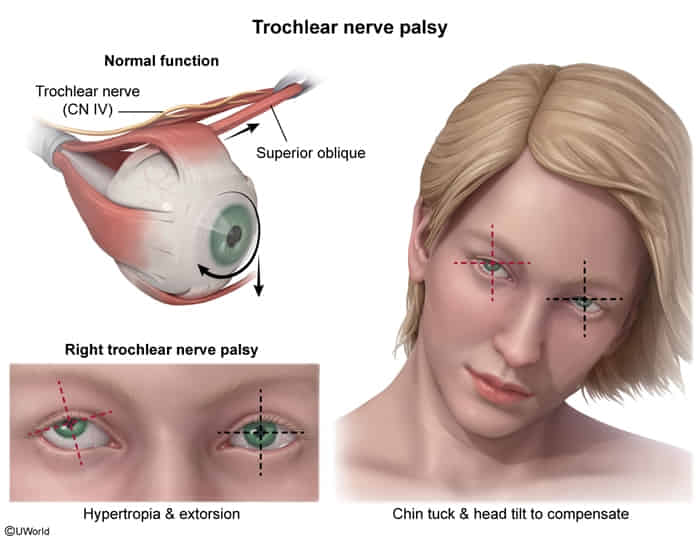
- Ocular Misalignment:
- Esotropia: Inward/nasal deviation (most common).
- Exotropia: Outward/temporal deviation.
- Hypertropia/Hypotropia: Vertical deviation (upward/downward).
- Compensatory behaviors: Head tilt, abnormal head posture, or squinting/closing one eye in bright sunlight.
- Visual symptoms:
- Young children: Suppression of image from the deviating eye (often asymptomatic, but leads to visual loss).
- Older children / Adults: Diplopia (double vision) or asthenopia (eye strain).
Diagnosis
- Initial / Bedside Screening:
- Corneal light reflex test (Hirschberg test): Asymmetric deflection of light off the corneas (normal = symmetric, central reflex).
- Cover test / Cover-uncover test:
- Cover test: Detects manifest deviation (tropia). The uncovered deviating eye shifts to fixate when the straight eye is covered.
- Cover-uncover test: Detects latent deviation (phoria).
- Essential Workup:
- Cycloplegic refraction: To assess for uncorrected refractive errors (hyperopia).
- Dilated funduscopic exam: Crucial to rule out underlying intraocular pathology (e.g., retinoblastoma, optic nerve hypoplasia). c
- Imaging (MRI/CT Orbit & Brain):
- Indicated if: Sudden/acute onset, paralytic/incomitant strabismus (CN III, IV, or VI palsy), presence of neurologic signs, papilledema, or suspected retinoblastoma/intracranial neoplasm.
Differential Diagnostics
- Pseudostrabismus:
- Diff: False appearance of esotropia due to prominent epicanthal folds or a broad, flat nasal bridge. Shows symmetric corneal light reflex and normal cover test.
- Retinoblastoma:
- Diff: Manifests with leukocoria (white pupillary reflex) and intraocular mass visible on fundoscopic exam / ocular US / MRI.
- Cranial Nerve Palsy (CN III, IV, VI):
- Diff: Sudden onset, limited extraocular motility (incomitant deviation depending on direction of gaze). Often associated with trauma, ↑ ICP, ischemia, or neoplasm.
- Congenital Cataract:
- Diff: Leukocoria, absent or abnormal red reflex; visual deprivation leads to secondary sensory strabismus.
Management
Primary goal: Prevent irreversible amblyopia and preserve binocular vision (depth perception).
- First-Line / Conservative:
- Refractive Correction: Prescribe corrective lenses (glasses) (curative for accommodative esotropia driven by hyperopia).
- Occlusion Therapy (for Amblyopia):
- Patching the sound/dominant eye for several hours daily to force stimulation of the amblyopic eye.
- Pharmacologic Penalization: Topical atropine drops placed in the sound eye (blurs near vision, forcing use of the amblyopic eye; alternative if poor patch adherence).
- Second-Line / Surgical:
- Extraocular Muscle (EOM) Surgery (recession/resection):
- Indicated for: Congenital (infantile) esotropia, persistent deviation despite full refractive/patching therapy, or large non-accommodative deviations.
- Optimal timing: Usually performed early (prior to age 1–2) for congenital esotropia to maximize binocular fusion.
- Extraocular Muscle (EOM) Surgery (recession/resection):
- Refractory / Alternative:
- Botulinum toxin (Botox) injection: Direct injection into the overacting EOM (temporary realignment or adjunct in select cases).
Complications
- Loss of Stereopsis: Deficits in binocular depth perception.
- Contracture of Extraocular Muscles: Chronic misalignment causing mechanical shortening of the affected rectus muscle.
- Psychosocial and cosmetic impairment.
Amblyopia
- Pathophysiology: Functional ↓ visual acuity (VA) due to abnormal visual cortex development and image suppression during early childhood (< 7–8 yo).
- Etiologies:
- Strabismic (most common): Ocular misalignment (esotropia/exotropia) causes suppression of the deviated image.
- Refractive / Anisometropic: Unequal refractive error between eyes; visual cortex favors the clearer image. c
- Deprivational (most severe): Physical occlusion of visual axis (e.g., congenital cataract, severe ptosis, retinoblastoma).
- Clinical Features:
- Typically asymptomatic; detected via routine screening.
- Squinting, head-tilting, bumping into objects, poor stereopsis (depth perception).
- PE: Asymmetric VA (≥ 2-line difference on chart), abnormal corneal light reflex (Hirschberg), or abnormal red reflex (Brückner).
- Diagnosis:
- Screening: Red reflex in infants; formal VA testing (LEA symbols, Snellen) starting at age 3–5.
- Workup: Cycloplegic refraction (detects refractive errors) + dilated fundoscopy (rules out retinoblastoma, cataract, retinal pathology).
- Criteria: ↓ VA not immediately correctable by refraction alone, in the absence of structural retinal/optic nerve defects.
- Management (critical before age 7–8 due to visual neuroplasticity):
- Initial: Treat primary etiology (urgent cataract removal; corrective lenses for refractive error).
- First-line: Occlusion therapy (patching) of the sound (better-seeing) eye for 2–6 hrs/day.
- Alternative / Second-line: Atropine 1% drops instilled in the sound eye (blurs near vision; preferred if poor patch compliance).
- Refractory / Residual strabismus: Extraocular muscle surgery after amblyopia is maximally treated.
- Complications:
- Irreversible vision loss / blindness in the amblyopic eye if untreated past critical window (> 8 yo).
- Reverse (occlusion) amblyopia in the sound eye due to excessive patching or atropine use.