Etiology
- Primary HPTH:
- Peak incidence 50-60yo; F:M ratio is 3:1.
- Risk factors: Radiation exposure to neck, chronic lithium use.
- Associated with MEN1 (pituitary, pancreatic, parathyroid) and MEN2A (pheochromocytoma, medullary thyroid cancer, parathyroid).
- Secondary HPTH:
- Chronic Kidney Disease (CKD) is the most common cause (impaired phosphate excretion and ↓ 1,25-(OH)2D3).
- Malabsorption (Vitamin D deficiency).
- Tertiary HPTH:
- Long-standing CKD leading to autonomous parathyroid hyperplasia, typically post-renal transplant. c
- Due to downregulation of calcium-sensing receptor and vitamin D receptor in the parathyroid glands
- Long-standing CKD leading to autonomous parathyroid hyperplasia, typically post-renal transplant. c
Parathyroid function
- Bone: Increases bone resorption by indirectly activating osteoclasts (via RANKL on osteoblasts), releasing Ca2+ and PO4(3-).
- Kidney: Increases Ca2+ reabsorption in the distal convoluted tubule. Decreases PO4(3-) reabsorption in the proximal convoluted tubule (“Phosphate trashing”). Stimulates 1-alpha-hydroxylase to convert 25-hydroxyvitamin D to active 1,25-dihydroxyvitamin D (calcitriol).
- Gut: Active Vitamin D increases intestinal absorption of both Ca2+ and PO4(3-).
Clinical features
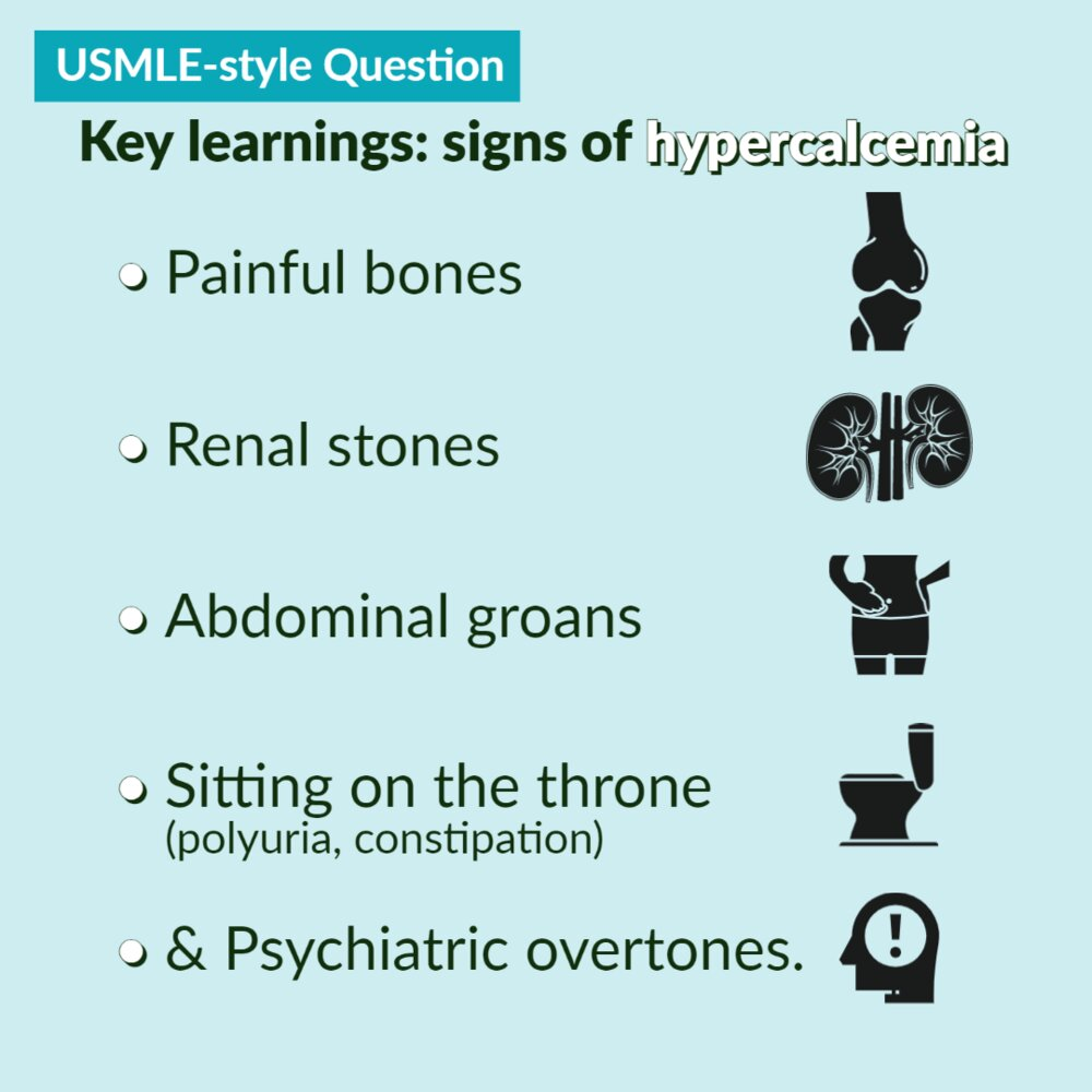
Symptomatic patients often have clinical features of hypercalcemia
Clinical features
- “Stones”: Nephrolithiasis, nephrocalcinosis.
- “Bones”: Bone pain, pathological fractures, osteitis fibrosa cystica (subperiosteal bone resorption in PHPT).
- “Groans”: Constipation, nausea, vomiting, anorexia, abdominal pain, pancreatitis.
- “Thrones”: Polyuria, polydipsia (due to nephrogenic diabetes insipidus). c
- Due to acquired renal ADH resistance. Although ADH is being secreted, the kidneys no longer respond to it adequately (nephrogenic diabetes insipidus).
- Physiological teleology: Stone Prevention: If the kidney concentrated urine in the setting of hypercalciuria (high urinary Ca), the calcium would precipitate into stones (nephrolithiasis)
- “Psychiatric overtones”: Fatigue, depression, confusion, lethargy, stupor/coma.
- Cardiac: Bradycardia, AV block, shortened QT interval on ECG.
Link to original
Diagnostics
- Labs
- Primary hyperparathyroidism: ↑ Serum Ca2+, ↑ PTH, ↓ Serum PO₄³⁻, ↑ Urinary Ca2+, ↑ Urinary cAMP
- The severe hypercalcemia overwhelms the kidney’s ability to reabsorb calcium, resulting in a net increase in urinary calcium (hypercalciuria). c
- PTH acts on the kidney via a Gs-coupled receptor, which increases cAMP. This cAMP is then excreted, leading to ↑ Urinary cAMP.
- Primary hyperparathyroidism: ↑ Serum Ca2+, ↑ PTH, ↓ Serum PO₄³⁻, ↑ Urinary Ca2+, ↑ Urinary cAMP
- Neck imaging
- For surgical planning to determine the location of the abnormal glands and evaluate for concomitant thyroid disease
- Options include ultrasound neck and nuclear imaging, i.e., Tc-99m sestamibi scan.
- X-ray
- Decreased bone mineral density
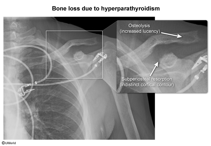
- Cortical thinning: especially prominent in the phalanges of the hand; manifests as acroosteolysis (a subperiosteal pattern of bone resorption)
- Unlike the typical osteoporosis of aging, which predominantly affects trabecular bone
- Salt and pepper skull: granular decalcification manifesting as diffusely distributed lytic foci on imaging of the calvarium
- Features of osteitis fibrosa cystica
Complications
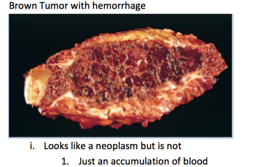
Osteitis fibrosa cystica (OFC)
- A rare skeletal disorder seen in advanced hyperparathyroidism characterized by replacement of calcified bone with fibrous tissue c
- Most commonly seen in primary hyperparathyroidism but can also occur in secondary hyperparathyroidism
- ↑ PTH → ↑ RANK ligand expression → activation of osteoclasts → bone resorption, cortical bone destruction, and fibrous tissue deposition
- Features include bone pain, subperiosteal thinning, and bone cysts; multiple bone cysts in the skull may result in a salt and pepper skull (pepper pot) appearance on x-ray.
- In advanced OFC, large, cystic, vascular cavities with a tumor-like appearance on x-ray and a brown color due to hemosiderin deposition (brown tumors) can form in long bones.