Epidemiology
- Most common cause of hyperthyroidism in the United States
Etiology
- Genetic predisposition
- 50% of patients with Graves disease have a family history of autoimmune disorders (e.g., type 1 diabetes mellitus, Hashimoto disease, pernicious anemia, myasthenia gravis)
- Associated with HLA-DR3 and HLA-B8 alleles
- Autoimmunity: B and T lymphocyte-mediated disorder
- Triggers
- Infectious agents: Yersinia enterocolitica and Borrelia burgdorferi have been shown to trigger antigen mimicry for homologies between their protein constituents and thyroid autoantigens.
- Stress
- Stress causes cortisol-mediated immune suppression, which is often followed by rebound immunologic hyperactivity and can trigger Graves disease in genetically susceptible individuals.
- Physical: surgery, trauma
- Psychological
- Pregnancy
- Thyroid overstimulation by high levels of hCG and TRAb during the first trimester increases the risk of Graves disease in the first trimester of pregnancy. In the second trimester, the risk decreases due to progesterone-induced immunosuppression. The risk of Graves disease increases postpartum in susceptible patients due to a drop in progesterone level which leads to the rebound of the immune system that can trigger autoimmunity.
Pathophysiology
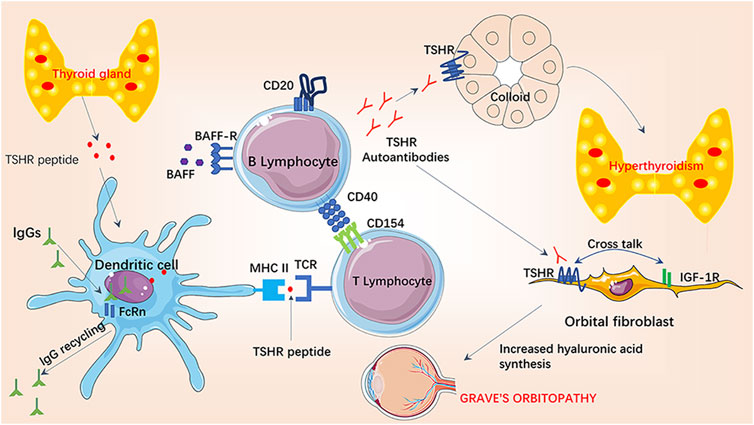
- General mechanism: B and T cell-mediated autoimmunity → production of stimulating immunoglobulin G (IgG) against TSH-receptor (TRAb; type II hypersensitivity reaction) → ↑ thyroid function and growth → hyperthyroidism and diffuse goiter
- Thyroid-associated ophthalmopathy
- TSH receptor is also present on fibroblasts in eyes
- Activated B and T cells infiltrate retro-orbital space targeting orbital fibroblasts → cytokine release (e.g. TNF-α, IFN-γ) → local inflammatory response → stimulates fibroblasts to secrete glycosaminoglycans (hyaluronic acid), which also pulls water into the interstitial space (osmotic effect) → expansion of retro-orbital tissue due to increased fluid in extraocular muscles, lymphocytic infiltration, and adipogenesis
- Pretibial myxedema: dermal fibroblast stimulation and deposition of glycosaminoglycans in connective tissue
Tip
Although the majority of symptoms are due to increased thyroid hormone levels, myxedema (nonpitting edema) and exophthalmos in patients with Graves disease are instead due to autoantibody-mediated activation of TSH receptors on peripheral tissue. Therefore they are not seen on other Hyperthyroidism diseases.
Clinical features
Diagnostics
Pathology
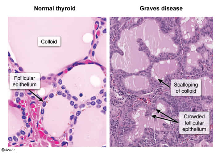
- Microscopic: histological features of an overactive gland
- Diffuse hyperplasia of thyroid follicles
- Tall, hyperplastic and hypertrophic follicular cells
- Hyperactive reabsorption causes scalloping around the edges of the colloid
Treatment
- Symptomatic (Immediate): Beta-blockers (Propranolol or Atenolol) for adrenergic symptoms.
- Definitive Therapies:
- Antithyroid Drugs (ATDs):
- Methimazole (MMI): 1st-line for most pts. Teratogenic in 1st trimester (aplasia cutis).
- PTU: 1st-line in 1st trimester of pregnancy & thyroid storm (blocks peripheral T4 -> T3). Hepatotoxic.
- Adverse Effect: Agranulocytosis -> discontinue drug and obtain urgent CBC if fever/sore throat occurs.
- Radioactive Iodine (RAI / I-131):
- Destroys follicular cells -> lifelong levothyroxine.
- Absolute contraindications: Pregnancy and lactation.
- Caution: Can worsen orbitopathy (pre-treat with prednisone if mild-moderate). c
- possibly due to an increase in TRAB levels caused by leakage of thyroid antigens from the irradiated thyroid
- Total Thyroidectomy:
- Indications: Compressive goiter, coexisting malignancy, severe active orbitopathy.
- Pre-op prep: Euthyroid on ATDs + potassium iodide (Lugol solution) to ↓ vascularity.
- Antithyroid Drugs (ATDs):
- Graves ophthalmopathy
- High-dose IV steroids
- Glucocorticoids decrease the severity of inflammation and reduce the excess extraocular volume.
- High-dose IV steroids
Special patient groups
Neonatal Graves disease
- Pathophysiology: Transplacental passage of maternal TSH-receptor antibodies (TRAb/TSI) (IgG class). Can occur even if mother is post-thyroidectomy/ablation.
- Clinical Presentation:
- Tachycardia, hypertension, irritability, tremors.
- Poor weight gain / FTT despite voracious appetite, diarrhea.
- Diffuse goiter (risk of airway obstruction), exophthalmos, warm skin.
- Onset: Delayed (7-10 days post-birth) if mother took antithyroid drugs (ATDs) near term, as maternal ATDs clear neonatal circulation faster than TRAb.
- Diagnosis:
- ↑ Free T4 & T3, ↓ TSH.
- Detection of maternal/neonatal TRAb.
- Management:
- Course: Transient; resolves spontaneously within 3-12 weeks as maternal IgG clears.
- Mild/Asymptomatic: Observation.
- Symptomatic/Severe: Methimazole (inhibits synthesis) + Propranolol (controls tachycardia/adrenergic symptoms).
- Refractory/Heart Failure: Add Lugol’s solution (potassium iodide; acutely blocks release) +/- hydrocortisone (decreases peripheral T4 to T3 conversion).
- Key Complication: High-output heart failure and craniosynostosis.