Epidemiology
Peak incidence: 10–30 years
Etiology
- Autoimmune destruction of epidermal melanocytes.
- Peak onset: Age 10–30 yrs; equal prevalence across sexes and ethnicities (cosmetically more evident in darker skin types).
- Strong association with other autoimmune diseases:
- Autoimmune thyroid disease (Hashimoto thyroiditis/Graves dz — most common association; ~20–30% of pts). c
- Type 1 Diabetes Mellitus (T1DM).
- Alopecia areata.
- Pernicious anemia (atrophic gastritis).
- Primary adrenal insufficiency (Addison disease).
- Family history present in ~30% of affected individuals.
Pathophysiology
Clinical features
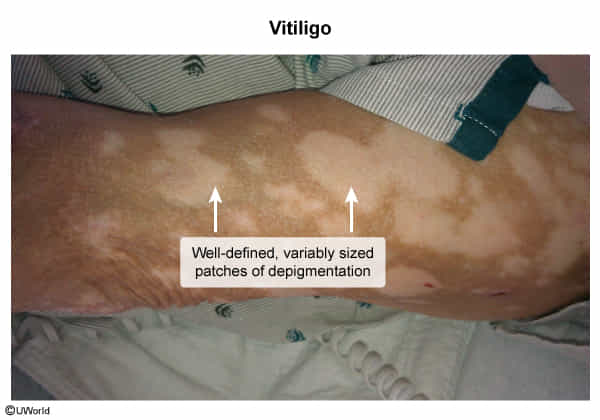
- Irregular, well-demarcated, depigmented (white) macules or patches
- Unilateral or bilateral distribution, with stable or slowly progressive lesions
- Leukotrichia: depigmented hair; an indicator of poor prognosis
- Coexisting autoimmune diseases (∼ 20% of patients)
- Most common: Hashimoto thyroiditis, Graves disease
- Other disorders: type 1 diabetes, psoriasis, inflammatory bowel disease, alopecia areata, pernicious anemia, Addison disease
Diagnostics
- Initial / Best Step: Clinical diagnosis based on history, pattern recognition, and physical examination.
- Diagnostic Modalities:
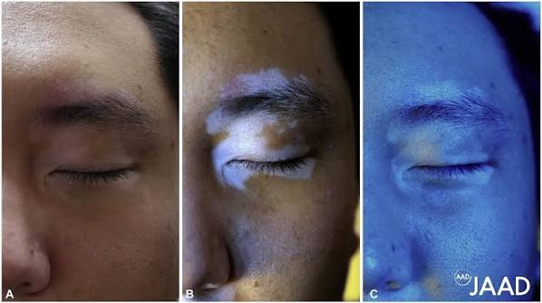
- Wood’s lamp examination: Highlights lesions with a bright, chalky-white/blue fluorescence (sharpens contrast against normal surrounding skin; useful in fair-skinned individuals).
- Key Labs: Screening for associated autoimmune endocrinopathies:
- Serum TSH (most important screening test; check anti-TPO if abnormal). c
- Fasting blood glucose or HbA1c.
- Complete blood count (CBC) to screen for pernicious anemia.
- Skin Biopsy (Rarely indicated):
- Performed only if diagnosis is ambiguous.
- Histology: Total absence of melanocytes in the basal layer (confirmed via Fontana-Masson or Melan-A staining) and loss of epidermal melanin pigment.
- Wood’s lamp examination: Highlights lesions with a bright, chalky-white/blue fluorescence (sharpens contrast against normal surrounding skin; useful in fair-skinned individuals).
- Differential diagnostics: see Catecholamine Synthesis > Albinism