Epidemiology
- Demographics: Bimodal peak (15–35 yrs and 55–70 yrs); equal ♂:♀ distribution.
- Genetics: Strong familial link; strongly associated with HLA-Cw6.
- Triggers & Risk Factors:
- Physical trauma: Friction, scratching, burns (Koebner phenomenon).
- Infections: Group A Streptococcus pharyngitis (triggers guttate psoriasis); HIV infection (triggers sudden, severe, or treatment-refractory disease). c
- Medications: Beta-blockers, Lithium, Antimalarials, NSAIDs, and systemic corticosteroid withdrawal (triggers pustular/erythrodermic flare).
- Lifestyle/Environment: Stress, smoking, heavy alcohol intake, cold/dry weather.
Pathophysiology
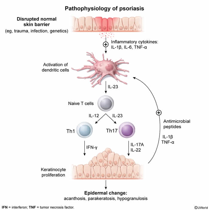
- Increased proliferation of keratinocytes
- Normally, the epidermis takes about 26–28 days to completely renew itself. This process involves the formation of new keratinocytes and shedding of aged (keratotic) skin cells. In individuals with psoriasis, the proliferation rate increases five-fold to ten-fold.
- Acanthosis: thickening of the epidermis = typical erythematous plaques
- Parakeratosis: retention of nucleated keratinocytes in the stratum corneum = characteristic silvery scales
- T cells secrete cytokines, which mediate an inflammatory response.
Clinical features
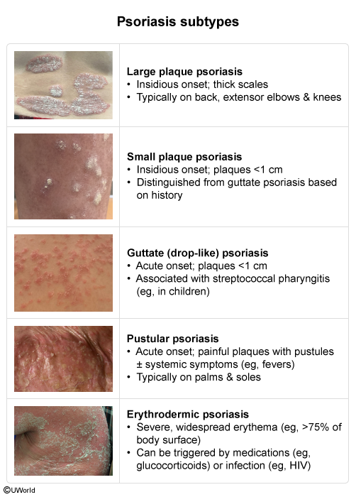
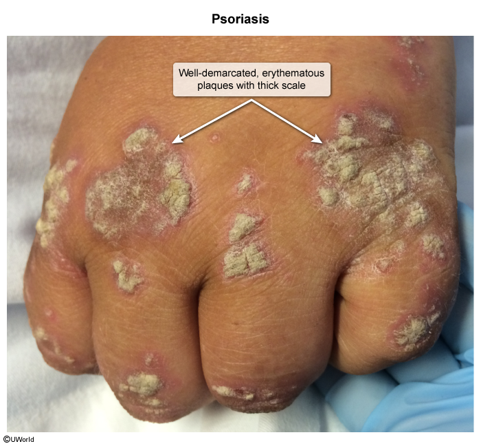
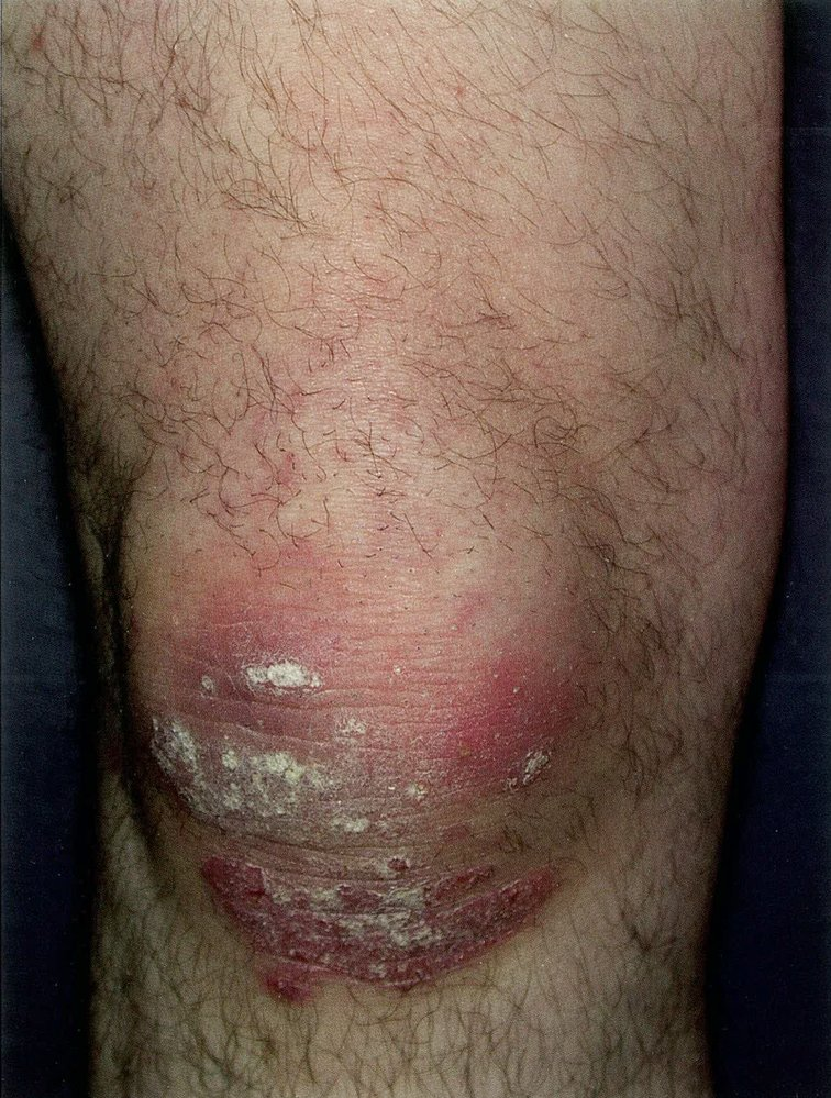
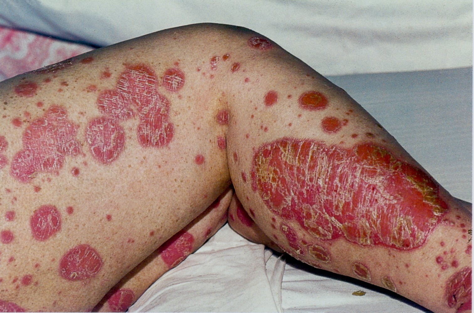
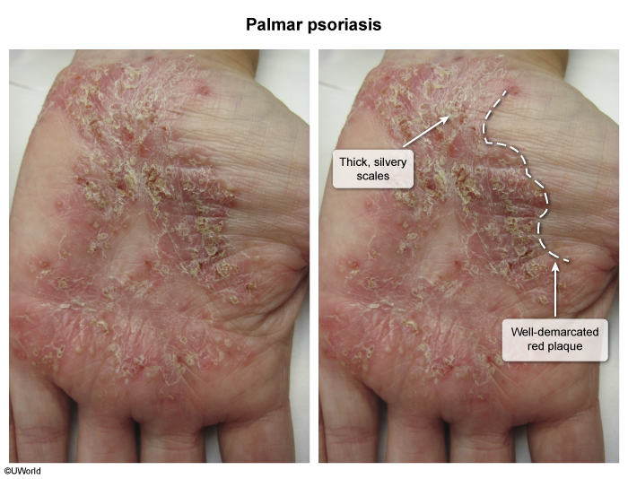
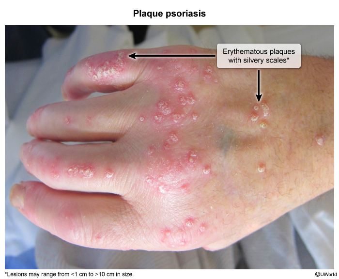
- Plaque Psoriasis (Psoriasis Vulgaris): ~80–90% of cases.
- Well-demarcated, erythematous plaques covered by coarse, silvery-white scale.
- Predilection for extensor surfaces (elbows, knees), scalp, lumbosacral region, and umbilicus.
- Auspitz sign: Punctate bleeding spots upon scraping off the scale (due to dilated dermal capillaries).
- Koebner phenomenon: Development of psoriatic lesions at sites of local skin injury.
- Guttate Psoriasis:
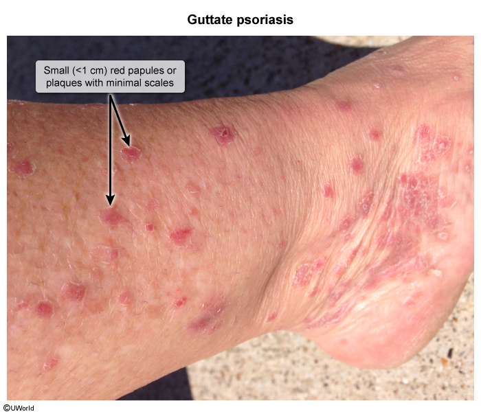
- Acute eruption of numerous small (2–10 mm), teardrop-shaped papules/plaques on the trunk and proximal extremities.
- Classically occurs 1–3 weeks after streptococcal pharyngitis in children/young adults.
- Pustular Psoriasis:
- Acute eruption of generalized, sterile, coalescing pustules on an erythematous base.
- Systemic signs: High fever, leukocytosis, electrolyte derangements. Often precipitated by rapid taper of systemic steroids.
- Erythrodermic Psoriasis:
- Diffuse erythema and scaling affecting >90% of total BSA.
- Dermatologic emergency: Causes impaired thermoregulation, fluid/protein loss, and high-output heart failure.
- Inverse Psoriasis:
- Smooth, well-demarcated, erythematous plaques lacking scale, located within intertriginous folds (axillae, inguinal creases, inframammary).
- Nail Involvement: Seen in up to 50% of skin disease and >80% of psoriatic arthritis (PsA).
- Nail pitting, “oil-drop” discoloration (salmon patches), subungual hyperkeratosis, and onycholysis.
Associated Conditions
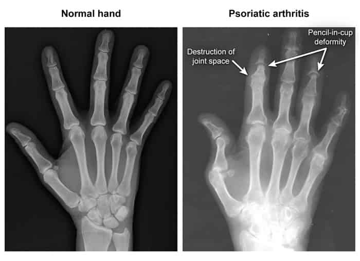
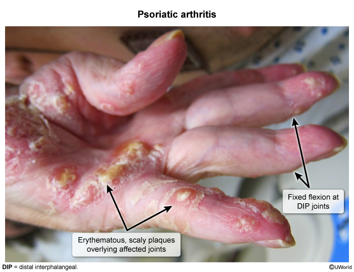
- Psoriatic arthritis (10-30%): asymmetric oligoarthritis, DIP involvement, “sausage digits” (dactylitis), pencil-in-cup deformity on X-ray t

- Metabolic syndrome, cardiovascular disease, IBD
Diagnostics
Skin biopsy
- Indications: Consider only if clinical presentation is atypical.
- Supportive findings
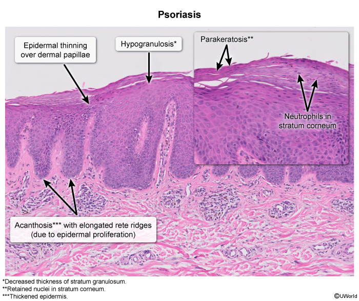
- Acanthosis and parakeratosis
- Thickening of the stratum spinosum, thinning of the stratum granulosum
- Acanthosis = typical erythematous plaques
- Parakeratosis = characteristic silvery scales
- Munro microabscesses: accumulation of neutrophils in the stratum corneum surrounded by parakeratosis
Differential diagnostics
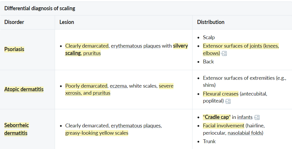 vs
vs 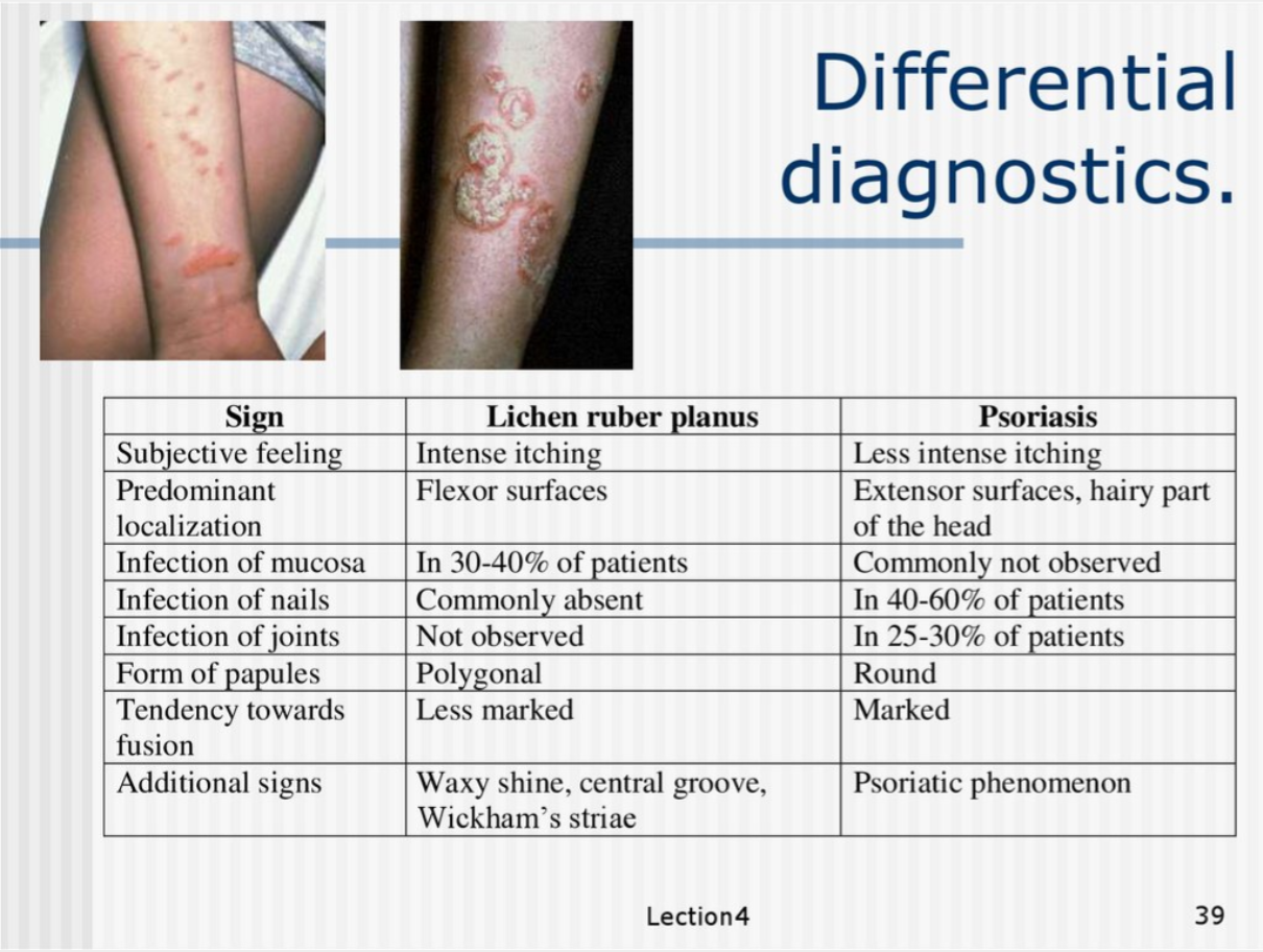
Psoriasis vs Lichen planus
- Immune Mechanism:
- Lichen Planus primarily involves CD8+ T cells targeting basal keratinocytes, creating a band-like lymphocytic infiltrate at the dermo-epidermal junction and interface dermatitis
- Psoriasis is driven by Th17/Th1 cells and involves complex cytokine cascades (IL-17, IL-22, TNF-α), affecting the entire epidermis and leading to neutrophil recruitment
- Keratinocyte Response:
- Lichen Planus shows normal to slow keratinocyte turnover, allowing time for proper differentiation, resulting in hypergranulosis (increased keratohyalin granules)
- Because of immune stress?
- Psoriasis exhibits extremely rapid keratinocyte turnover (3-5 days vs normal 28-30 days), preventing proper differentiation, leading to loss of granular layer and parakeratosis
Treatment
- Mild-Moderate Disease:
- Topical Corticosteroids (High-potency first-line).
- Vitamin D Analogs (e.g., Calcipotriene). t
- Alternatives: Tazarotene (retinoid), Tacrolimus (face/groin).
- Moderate-Severe/Refractory:
- Phototherapy: Narrowband UVB (preferred) or PUVA.
- Systemic (Non-Biologic): Methotrexate t , Cyclosporine, Acitretin.
- Biologics: TNF-α inhibitors (Adalimumab), IL-17/IL-23 inhibitors (Ustekinumab, Secukinumab). Screen for TB first.
- Important Contraindication:
- NO Systemic Corticosteroids: Withdrawal can trigger pustular psoriasis.