- Patho/Etiology
- Structural defects in the posterior fossa, leading to downward displacement of cerebellar and/or brainstem structures through the foramen magnum.
- Believed to result from a mismatch between the size of the developing cerebellum and the volume of the posterior fossa.
- Obstruction of normal cerebrospinal fluid (CSF) flow is a major cause of symptoms.
- Types & Presentation
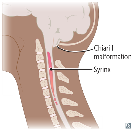
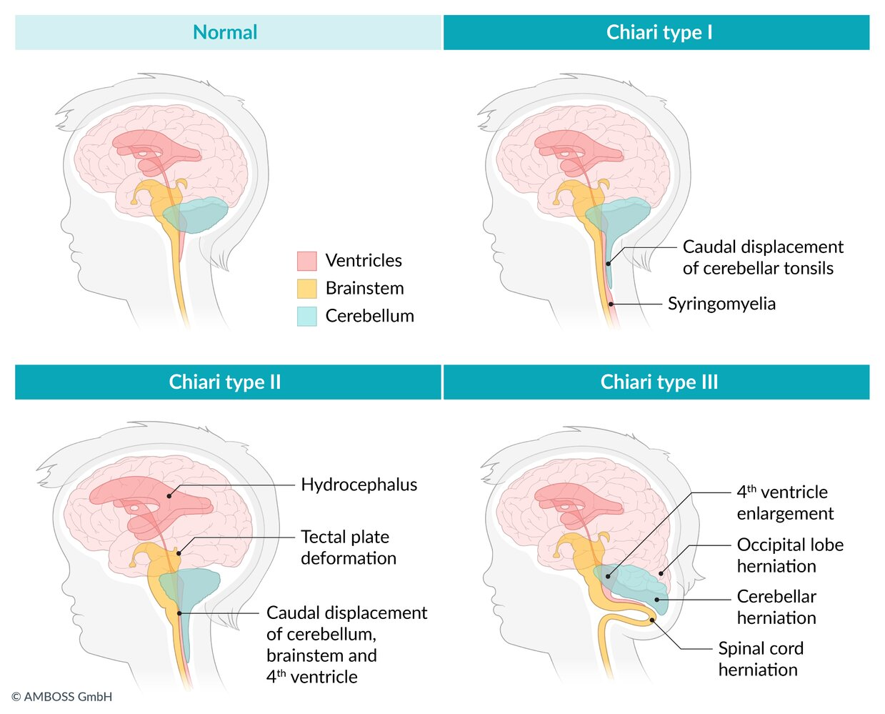
- Chiari I Malformation (Usually asymptomatic in childhood, manifests in adulthood)
- Anatomy: Low-lying cerebellar tonsils (>5 mm below foramen magnum).

- Presentation: Often asymptomatic in childhood, presents in adolescence/adulthood.
- Occipital headache and neck pain, classically exacerbated by coughing or Valsalva maneuver.
- Cerebellar symptoms (e.g., ataxia, nystagmus) and cranial nerve dysfunction (e.g., dysphagia, hoarseness).
- Key Association: Syringomyelia (fluid-filled cavity in the spinal cord), which can cause a “cape-like” distribution of lost pain and temperature sensation in the upper extremities.
- Anatomy: Low-lying cerebellar tonsils (>5 mm below foramen magnum).
- Chiari II Malformation (Arnold-Chiari Malformation) (More severe than Chiari I, usually presents early in life)
- Anatomy: Herniation of cerebellar vermis and tonsils, plus brainstem (medulla, pons) and a low-lying 4th ventricle through the foramen magnum.
- Presentation: Presents in infancy or early childhood.
- Symptoms are often due to brainstem compression and hydrocephalus, including stridor (a harsh vibrating noise when breathing), apnea, dysphagia, and limb weakness.
- Key Association: Almost universally associated with myelomeningocele (a severe form of spina bifida). Non-communicating hydrocephalus is also very common.
- Chiari I Malformation (Usually asymptomatic in childhood, manifests in adulthood)
- Diagnosis
- MRI is the gold standard imaging modality to visualize the herniated structures, assess for hydrocephalus, and identify a syrinx.
- Management
- Asymptomatic Chiari I patients are typically monitored.
- Symptomatic patients often require surgery.
- Posterior fossa decompression (craniectomy) is the most common procedure, often with a laminectomy of C1, to relieve pressure and restore CSF flow.
- A ventriculoperitoneal (VP) shunt may be necessary to treat associated hydrocephalus.

Chiari malformation vs Dandy-Walker malformation
| Feature | Chiari Malformation | Dandy-Walker Malformation |
|---|---|---|
| Pathophysiology | Cerebellum herniates through foramen magnum due to small posterior fossa. | Agenesis of cerebellar vermis allows 4th ventricle to enlarge into a cyst. |
| Posterior Fossa | Small / Normal | Enlarged |
| Key Abnormality | Tonsillar (Type I) or vermis + tonsillar (Type II) herniation. | Absent cerebellar vermis. |
| Hydrocephalus | Common, due to CSF outflow obstruction. | Very common, due to cystic 4th ventricle. |
| Key Associations | - Type I: Syringomyelia. - Type II: Myelomeningocele. | Hydrocephalus, Spina Bifida, Agenesis of corpus callosum. |
| Typical Presentation | - Type I: Adult w/ headaches. - Type II: Infant w/ brainstem dysfunction. | Infant w/ macrocephaly. |
Chiari畸形:过度拥挤的房间
请把后颅窝想象成一个小卧室,把小脑和脑干想象成里面的家具。
- 问题所在: 在Chiari畸形中,这个房间建得太小了,无法容纳所有它需要放置的家具。
- 后果: 为了把所有东西都塞进去,一部分家具(小脑扁桃体)被挤了出来,并从唯一的开口——门道(枕骨大孔)——伸了出去。
- 结果: 这些堵在门口的家具阻碍了交通(脑脊液流动),导致房间内压力积聚(脑积水),并影响到走廊的墙壁(脊髓空洞症)。
核心:Chiari是一个_拥挤_问题。
Dandy-Walker畸形:缺失的帐篷支柱
请把后颅窝想象成一个大帐篷,把小脑蚓部想象成它主要的中央支撑柱。
- 问题所在: 在Dandy-Walker畸形中,这根中央帐篷支柱缺失了(蚓部发育不全)。
- 后果: 没有了支柱的支撑,帐篷的顶部(第四脑室)无法正常成型。它会塌陷并被“雨水”(脑脊液)充满,形成一个巨大的、充满液体的囊肿。
- 结果: 这个不断膨胀的水囊肿将帐篷壁向外推(后颅窝扩大),并堵住了拉链出口(脑脊液流出道),将所有的水都困在里面,导致整个帐篷都肿胀起来(脑积水)。
核心:Dandy-Walker是一个_结构缺陷_问题。