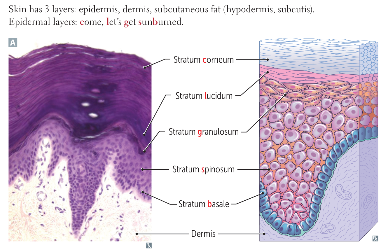
- Stratum corneum: outer layer of the epidermis
- Consists of dead (anuclear), keratin-filled cells
- This layer is constantly being sloughed off.
- Stratum lucidum: thin, translucent layer
- Located only on thick skin (palms and soles)
- Composed of a homogeneous layer of keratinocytes with no nuclei or organelles
- Stratum granulosum: also called the granular layer
- Contains keratohyalin
- This layer has waterproof properties.
- Stratum spinosum
- Composed of actively dividing keratinocytes with spinous-like projections (prickle cells)
- This layer produces keratin and induces keratinization.
- Langerhans cells are also located in this layer.
- If undergoes malignant changes, will lead to cSCC
- Stratum basale (also called the basal cell layer of the epidermis)
- Stem cells of the epidermis (their daughter cells migrate upwards and differentiate into other cells)
- Melanocytes and Merkel cells are also located in this layer.
- The stratum basale is regenerative (basal keratinocytes proliferate to fill skin defects).
Dermatopathology
Stratum Corneum
- Parakeratosis: Retention of nuclei in the stratum corneum.
- Classic Association: Psoriasis (due to rapid keratinocyte turnover).
- Also seen in: Actinic keratosis, Dandruff (Seborrheic dermatitis).
- Hyperkeratosis: Increased thickness of the stratum corneum.
- Classic Associations: Callus, Ichthyosis, Lichen Planus.
- Friction: Corns/calluses caused by repeated mechanical trauma.
- Disruption of Barrier:
- Atopic Dermatitis (Eczema): Defects in filaggrin (protein that aggregates keratin filaments) lead to barrier dysfunction and antigen entry.
Stratum Lucidum
- Note: USMLE rarely tests isolated pathology specific only to this layer, as it is strictly defined by location (Palms/Soles).
- Relevance: Involved in general Palmoplantar Keratodermas (thickening of palms/soles).
Stratum Granulosum
- Hypergranulosis: Increased thickness of the granular layer.
- Classic Association: Lichen Planus (Sawtooth infiltrate, Wickham striae).
- Hypogranulosis / Absent Granular Layer:
- Classic Association: Psoriasis (the layer thins or disappears due to rapid turnover).
- Classic Association: Ichthyosis Vulgaris (loss-of-function mutation in filaggrin gene).
Stratum Spinosum
- Acanthosis: Diffuse epidermal hyperplasia (thickening of the spinosum).
- Classic Associations: Acanthosis Nigricans (insulin resistance, GI malignancy), Psoriasis.
- Spongiosis: Epidermal accumulation of edematous fluid in intercellular spaces.
- Classic Association: Eczematous Dermatitis (Type IV HSR).
- Acantholysis: Separation of epidermal cells (loss of cohesion).
- Classic Association: Pemphigus Vulgaris (IgG Ab against Desmoglein 1 & 3).
- Findings: Flaccid bullae, + Nikolsky sign, reticular IF pattern.
- Histology: “Row of tombstones” (basal layer remains attached, spinosum detaches).
- Malignancy:
- Squamous Cell Carcinoma (cSCC): Malignant proliferation of keratinocytes with “keratin pearls.”
Stratum Basale
- Hemidesmosome Dysfunction:
- Classic Association: Bullous Pemphigoid (IgG Ab against Hemidesmosomes / BP180 & BP230).
- Findings: Tense bullae, - Nikolsky sign, linear IF pattern along BMZ.
- Differentiation: Entire epidermis lifts off the dermis (subepidermal blister).
- Melanocyte Pathology:
- Vitiligo: Autoimmune destruction of melanocytes.
- Albinism: Normal melanocyte number, ↓ Melanin production (Tyrosinase deficiency).
- Melanoma: Malignancy of melanocytes (S-100 tumor marker).
- Malignancy:
- Basal Cell Carcinoma (BCC): Most common skin cancer.
- Histology: Nests of “palisading” nuclei.