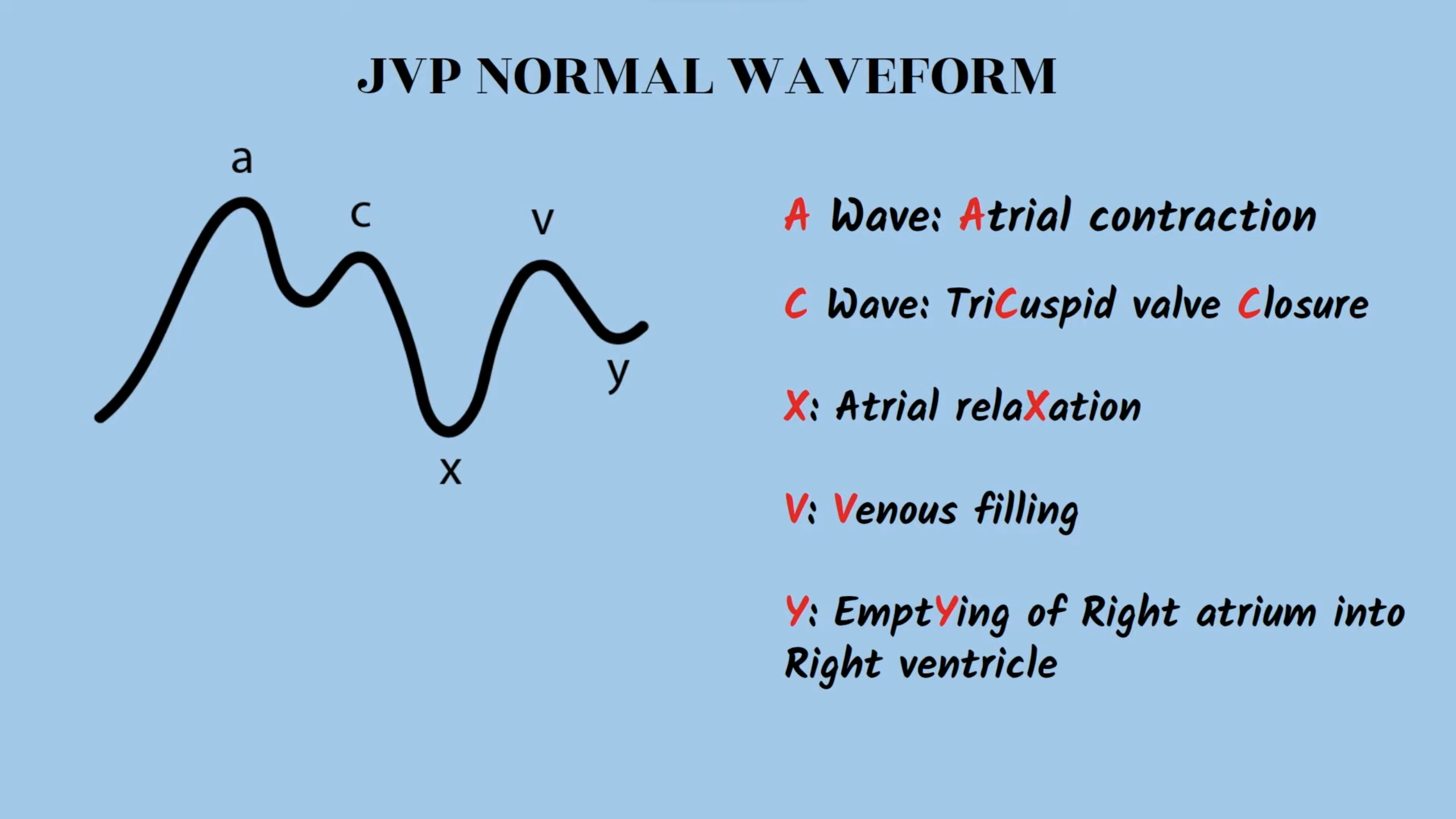
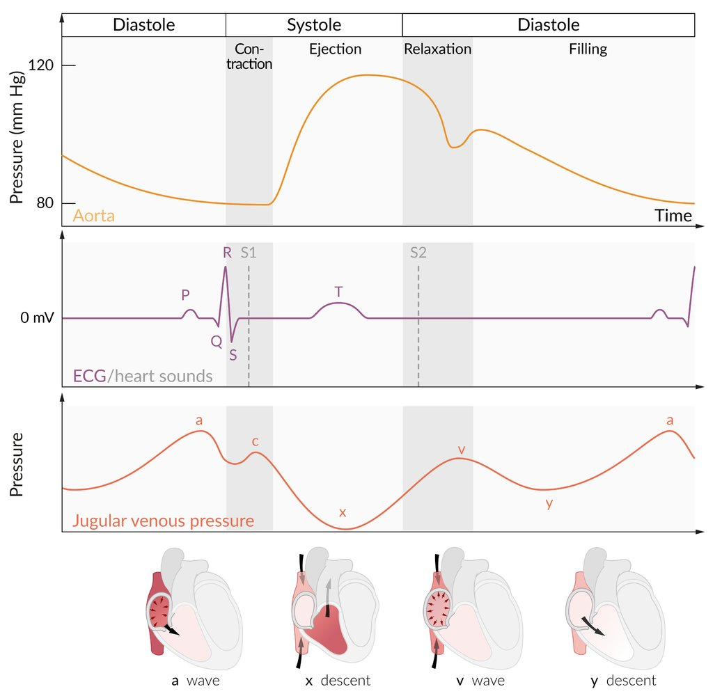
Normal Waveform Components
Abnormal JVP Waveforms
- Large ‘a’ wave: Increased resistance to right atrial emptying.
- Causes: Tricuspid stenosis, right ventricular hypertrophy, pulmonary hypertension.
- Cannon ‘a’ waves: Very large, intermittent ‘a’ waves.
- Pathophysiology: Right atrium contracts against a closed tricuspid valve (AV dissociation).
- Causes: Complete heart block (third-degree), ventricular tachycardia, premature ventricular/junctional contractions.
- Absent ‘a’ wave: No coordinated atrial contraction.
- Cause: Atrial fibrillation.
- Large ‘v’ wave (or c-v fusion wave):
- Pathophysiology: Blood regurgitates into the right atrium during ventricular systole.
- Cause: Tricuspid regurgitation.
- Rapid/Steep ‘y’ descent (Friedreich’s sign):
- Pathophysiology: Rapid, early diastolic filling of a stiff or non-compliant ventricle.
- Causes: Constrictive pericarditis, restrictive cardiomyopathy.
- Slow ‘y’ descent:
- Pathophysiology: Obstruction of right ventricular filling.
- Causes: Tricuspid stenosis, right atrial myxoma.
- Blunted/Absent ‘y’ descent:
- Pathophysiology: Impaired right ventricular filling due to external pressure.
- Cause: Cardiac tamponade.
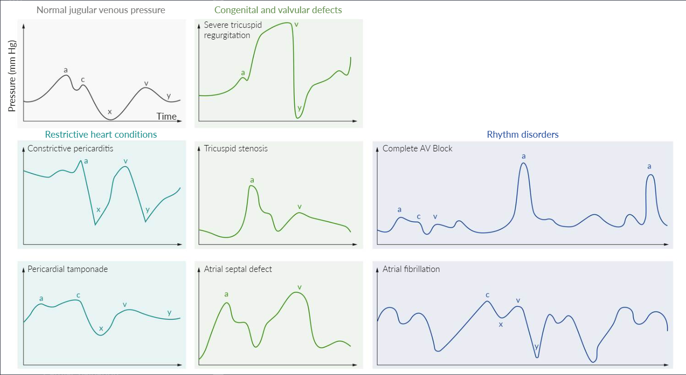
Pathology
Common abnormalities of the JVP waveform include:
- Constrictive pericarditis: elevated JVP (due to increased external atrial pressure) with a prominent x (exaggerated atrial relaxation) and y (early rapid ventricular filling) descent
- Cardiac tamponade: elevated JVP (due to increased external atrial pressure), a prominent x descent (exaggerated atrial relaxation), and a blunt or absent y descent (minimal ventricular filling)
- Tricuspid regurgitation: prominent v wave as the blood from the right ventricle regurgitates into the right atrium during ventricular systole (atrial diastole), increasing interatrial pressure and volume
- Tricuspid stenosis: giant a wave due to high right atrial systolic pressure
- Atrial septal defect: v wave ≥ a wave due to the left-to-right shunting of blood
- Third-degree atrioventricular (AV) block: cannon a waves due to the loss of AV synchronization and contraction of the atria against a closed tricuspid valve
- Atrial fibrillation: absent a waves due to ineffective contraction of the atria