Definition: Serum total calcium >10.5 mg/dL. Remember to correct for albumin: Corrected Ca2+ = Measured Ca2+ + 0.8 * (4 - measured albumin).
Tip
- Total protein normal level 6.0 to 8.3 g/dL (60 to 83 g/L)
- Albumin normal level 3.4 to 5.4 g/dL (34 to 54 g/L)
- Phosphorus normal level 2.5 to 4.5 g/dL (25 to 45 g/L)
Etiology
- PTH-Dependent (High or inappropriately normal PTH)
- Primary Hyperparathyroidism: Most common outpatient cause. Usually due to a parathyroid adenoma.
- Familial Hypocalciuric Hypercalcemia (FHH): Autosomal dominant mutation in the Ca2+-sensing receptor (CaSR). Leads to high PTH and high serum Ca2+, but low urine Ca2+.
- Tertiary Hyperparathyroidism: Seen in patients with chronic kidney disease where longstanding secondary hyperparathyroidism leads to autonomous parathyroid function.
- PTH-Independent (Low PTH)
- Malignancy: Most common inpatient cause.
- PTHrP (PTH-related peptide) mediated: Secreted by squamous cell carcinomas (lung, head, neck), renal, and breast cancer. Mimics PTH actions.
- Osteolytic lesions: Direct bone destruction from metastases (e.g., breast cancer) or multiple myeloma (release of local cytokines).
- Ectopic Vitamin D production: Granulomatous diseases (e.g., sarcoidosis, TB) and lymphomas can have macrophages that express 1α-hydroxylase, leading to excess active Vitamin D (Calcitriol).
- Vitamin D Toxicity: Over-ingestion of vitamin D supplements.
- Medications: Thiazide diuretics (increase renal Ca2+ reabsorption), lithium.
- Immobilization: Increased osteoclast activity.
- Milk-alkali syndrome: Excessive intake of calcium and absorbable alkali.
- Malignancy: Most common inpatient cause.
Clinical features
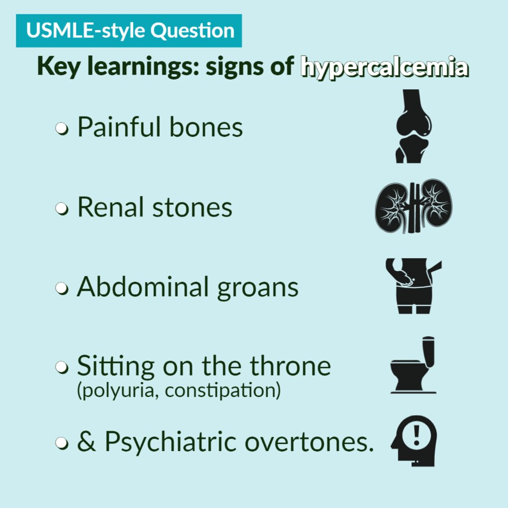
- Nephrolithiasis, nephrocalcinosis (calcium oxalate > calcium phosphate stones)
- Bone pain, arthralgias, myalgias, fractures
- Because most of the calcium is released from bones
- Constipation
- Increase in extracellular Ca2+ → membrane potential outside is more positive → more amount of depolarization is needed to initiate action potential → decreased excitability of muscle and nerve tissue
- Abdominal pain
- Nausea and vomiting
- Anorexia
- Peptic ulcer disease
- hypercalcemia-induced increase of gastric acid secretion and gastrin levels.
- Neuropsychiatric symptoms such as anxiety, depression, fatigue, and cognitive dysfunction
- Diminished muscle excitability
- Cardiac arrhythmias
- ECG: Shorten QT interval, see QT interval
- Muscle weakness, paresis
- Cardiac arrhythmias
- Polyuria and dehydration
- Due to acquired renal ADH resistance. Although ADH is being secreted, the kidneys no longer respond to it adequately (nephrogenic diabetes insipidus).

Diagnostics
- Step 1: Confirm hypercalcemia with a repeat measurement of total and ionized calcium, and correct for albumin.
- Step 2: Measure PTH level.
- High or inappropriately normal PTH → Suggests primary hyperparathyroidism or FHH.
- To differentiate, check 24-hour urine calcium. Low urine calcium suggests FHH; high or normal suggests primary hyperparathyroidism.
- Low PTH → Suggests malignancy or other non-PTH mediated causes.
- Workup includes measuring PTHrP, Vitamin D metabolites (25-OH and 1,25-(OH)2 D), SPEP/UPEP (for multiple myeloma), and imaging (e.g., chest X-ray) to search for malignancy.
- High or inappropriately normal PTH → Suggests primary hyperparathyroidism or FHH.
Treatment
- Consider calcitonin for rapid-onset, short-term control of hypercalcemia.
- Bisphosphonates for slow-onset, long-term control of hypercalcemia