Etiology
- Pathogens:
- Historically: Haemophilus influenzae type b (Hib) (most common in unvaccinated/undervaccinated).
- Current vaccinated era: Streptococcus pneumoniae, Streptococcus pyogenes (GAS), Staphylococcus aureus (including MRSA), non-typeable H. influenzae.
- Risk Groups:
- Unvaccinated/undervaccinated children (peak historically 2–7 years).
- Adults (mean age ~45 years, especially with DM, obesity, or immunosuppression).
Pathophysiology
Mnemonic
The hallmarks of epiglottitis are the three Ds: Dysphagia, Drooling, and Distress.
- Drooling is due to pain in swallowing.
Clinical features
- Acute, rapidly progressive course (<24 hours).
- The Classic “3 Ds”:
- Drooling (due to severe odynophagia).
- Dysphagia.
- Dysphonia (muffled, “hot potato” voice).
- Other Key Signs:
- High fever, severe sore throat out of proportion to pharyngeal exam (pharyngeal findings are typically normal) c
- Tripod/Sniffing position (sitting up, leaning forward, hyperextended neck, open mouth).
- Eases respiration as the airway diameter is increased by leaning forward and extending the neck in a seated position
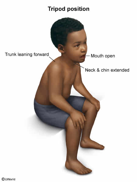
- Eases respiration as the airway diameter is increased by leaning forward and extending the neck in a seated position
- Inspiratory stridor, tachypnea, suprasternal/intercostal retractions.
- Absence of a barking cough (distinguishes from croup).
Diagnostics
- Initial/Screening:
- Clinical suspicion: Do NOT agitate the patient (avoid IV lines, supine positioning, or pharyngeal exam with tongue depressor prior to securing the airway risk of fatal laryngospasm).
- Imaging (Only if stable and diagnosis in doubt): c
- Lateral neck X-ray: Demonstrates “thumbprint sign” (swollen, enlarged epiglottis) and loss of the vallecular space.
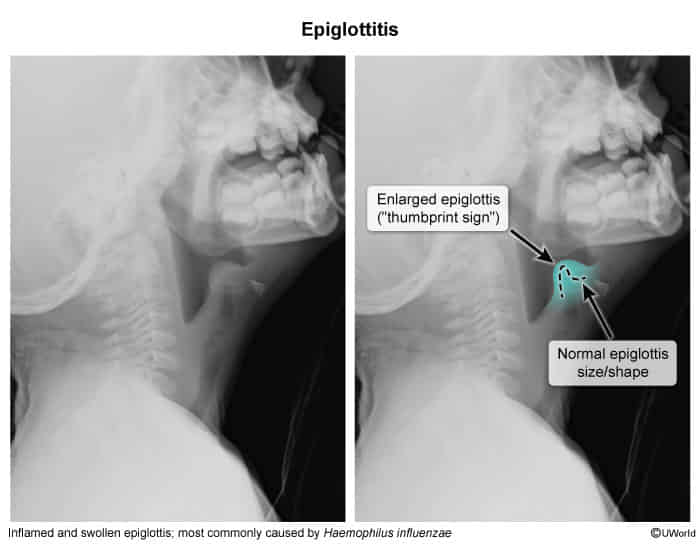
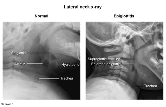
- Lateral neck X-ray: Demonstrates “thumbprint sign” (swollen, enlarged epiglottis) and loss of the vallecular space.
- Confirmatory/Gold Standard:
- Direct visualization/Nasopharyngolaryngoscopy: Performed strictly in a controlled setting (OR/ICU) with surgical airway backup.
- Shows swollen, cherry-red epiglottis and arytenoids.
- Key Labs (Obtained after airway is secured):
- Blood cultures (frequently positive in Hib).
- Epiglottic surface swab/culture.
- CBC: Marked leukocytosis with left shift.
Differential Diagnostics
- Croup (Laryngotracheobronchitis):
- Diff by viral prodrome, subacute onset, low-grade fever, “barking” seal-like cough, and “steeple sign” (subglottic narrowing) on AP neck X-ray. c
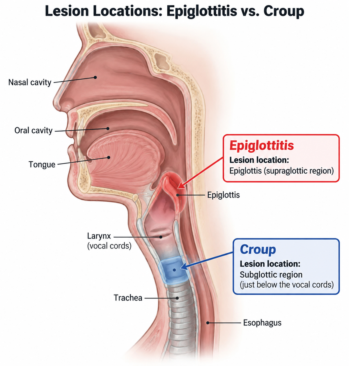
- Diff by viral prodrome, subacute onset, low-grade fever, “barking” seal-like cough, and “steeple sign” (subglottic narrowing) on AP neck X-ray. c
- Peritonsillar Abscess (Quinsy):
- Diff by unilateral tonsillar bulge, uvula deviation to contralateral side, and prominent trismus; common in older adolescents/young adults.
- Retropharyngeal Abscess:
- Diff by fever, limited neck extension/torticollis, and widened prevertebral soft tissue shadow on lateral neck X-ray; confirmed via contrast-enhanced CT neck.
- Foreign Body Aspiration:
- Diff by sudden-onset choking/stridor without fever or infectious prodrome; unilateral focal wheezing or air trapping on chest X-ray.
- Bacterial Tracheitis:
- Diff by “croup-like” illness that rapidly worsens with high fever, toxic appearance, and thick, purulent tracheal secretions resistant to nebulized epinephrine.
Treatment
- 1. Airway Stabilization (First-Line / Absolute Priority):
- Keep child calm (allow to remain in parent’s lap, provide blow-by ).
- Immediate controlled Endotracheal Intubation (ETT) in the operating room (OR) by skilled personnel (ENT and Anesthesia).
- Prepare for immediate surgical airway (cricothyrotomy/tracheostomy) if ETT fails.
- 2. Medical Therapy (Empiric IV Antibiotics):
- Initiate immediately after airway control:
- IV 3rd-generation cephalosporin (Ceftriaxone or Cefotaxime)
- PLUS an anti-staphylococcal agent covering MRSA (Vancomycin or Clindamycin).
- Duration: 7–10 days total (transition to PO based on sensitivities once extubated and afebrile).
- Initiate immediately after airway control:
- 3. Chemoprophylaxis:
- Rifampin prophylaxis for all household contacts if H. influenzae type b is isolated AND there is an unvaccinated/undervaccinated child yo or an immunocompromised contact in the home.