Epidemiology
Etiology
Predisposing factors
- Nulliparity
- Early menarche (< 10 years of age)
- Age: 25–45 years
- Fibroids are largely found in women of reproductive age.
- Influenced by hormones (i.e., estrogen, growth hormone, and progesterone)
- During menopause, hormone levels begin to decrease and leiomyomas begin to shrink.
- Increased incidence in African American individuals
Pathophysiology
Clinical features
- Symptoms depend on the number, size, and location of leiomyomas. Often asymptomatic (up to 80% of cases).
- Abnormal menstruation (possibly associated with anemia): hypermenorrhea, heavy menstrual bleeding, metrorrhagia, dysmenorrhea
- Submucosal leiomyomas are most frequently associated with significantly prolonged or heavy menstrual bleeding. The mechanism may be related to the increased total surface area as a result of the bulging uterine wall, impaired uterine wall contractility, or micro/macrovascular abnormalities.
- Features of mass effect
- Enlarged, firm and irregular uterus during bimanual pelvic examination
- Size can range from normal to full-term gestation
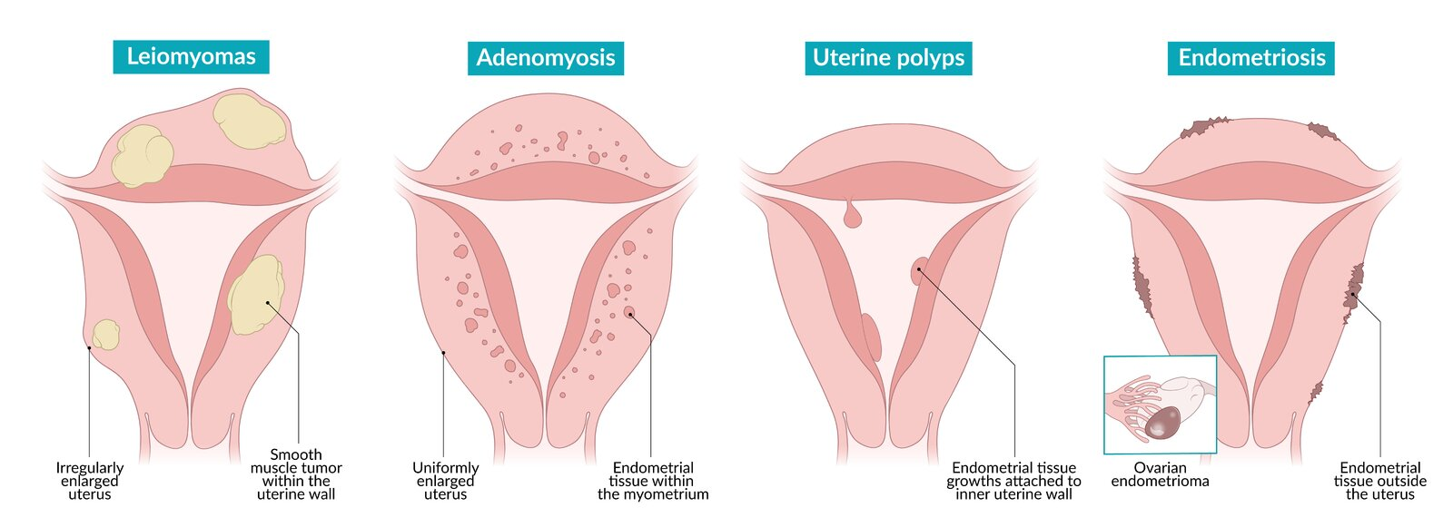
- Differ from Adenomyosis, which shows globular, uniformly enlarged uterus that is soft but tender on palpation
- Back or pelvic pain/discomfort
- Urinary tract or bowel symptoms (e.g., urinary frequency/retention/incontinence, constipation, features of hydronephrosis)
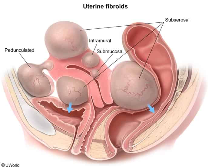
- Enlarged, firm and irregular uterus during bimanual pelvic examination
- Reproductive abnormalities
- Infertility (difficulty conceiving and increased risk of pregnancy loss), especially with submucosal fibroids.
- Related to an obstructed uterine cavity and/or impaired contractility of the uterus.
- Subserosal fibroids are located outside the uterine cavity and therefore typically do not interfere with implantation or cause miscarriage. c
- Dyspareunia
- Infertility (difficulty conceiving and increased risk of pregnancy loss), especially with submucosal fibroids.
Diagnostics
Warning
Uterine leiomyomas are extremely common (affecting 70% of women) and are often found incidentally on ultrasound; do not attribute abnormal uterine bleeding to leiomyomas until other etiologies have been ruled out!
Pathology
- Macroscopic
- Grayish-white surface
- Homogeneous; tissue bundles on cross-section partly in a whorled pattern
- Some leiomyomas may involve regressive changes: scar formation, calcification, and cysts
- Microscopic: Smooth muscle tissue in a whorled pattern with well-demarcated borders, consisting of monoclonal cells interspersed with connective tissue
Differential diagnostics
Uterine leiomyosarcoma (uterine sarcoma)
- Definition & Subtypes
- Aggressive mesenchymal uterine tumor.
- Leiomyosarcoma (LMS) is the most common subtype.
- Risk Factors
- Prior pelvic radiation (strongest environmental risk factor).
- Prolonged Tamoxifen use (> 5 yrs). c
- Postmenopausal age, African American race.
- Clinical Presentation
- Rapidly enlarging uterine mass in a postmenopausal woman.
- Abnormal uterine bleeding (AUB) / postmenopausal bleeding (PMB).
- Pelvic pain/pressure, bulk symptoms (urinary frequency, constipation).
- Diagnosis
- Initial: Pelvic US (heterogeneous mass, central necrosis, high vascular flow).
- Endometrial Biopsy: Often negative/non-diagnostic (tumor is intramural, not mucosal).
- Confirmatory: Surgical histopathology (post-hysterectomy).
- Staging: Chest CT (metastasis to lungs is #1).
- Management
- First-line: TAH-BSO (Total Abdominal Hysterectomy w/ Bilateral Salpingo-Oophorectomy).
- CRITICAL CONTRAINDICATION: Power morcellation is forbidden (causes peritoneal dissemination).
- Adjuvant Therapy: Chemotherapy (Doxorubicin-based) or RT for advanced LMS; progesterone/aromatase inhibitors for low-grade Endometrial Stromal Sarcoma (ESS).
- Differential Diagnosis
- Uterine Fibroid (Leiomyoma): Premenopausal, slow-growing, benign.
- Endometrial Carcinoma: Epithelial origin, readily diagnosed on endometrial biopsy.
Treatment
- Asymptomatic: Observation, as most fibroids shrink after menopause. c
- Medical (symptom control):
- Hormonal contraceptives (e.g., OCPs) or progestin-releasing IUDs can manage heavy bleeding but do not shrink fibroids.
- GnRH agonists (e.g., leuprolide) induce a temporary menopause-like state, shrinking fibroids. Used short-term pre-operatively due to significant side effects.
- Surgical/Procedural:
- Myomectomy: Surgical removal of fibroids while preserving the uterus. This is the choice for patients who desire future fertility.
- Hysterectomy: Definitive treatment, removing the uterus and thus curing the fibroids.
- Uterine Artery Embolization (UAE): A minimally invasive procedure where embolic agents block blood flow to the fibroids, causing them to shrink.
Special patient groups
Uterine leiomyomas during pregnancy
- Overview & Risk: Estrogen/progesterone-dependent; risk in AA women & advanced maternal age; presents as fundal height > GA.
- Red Degeneration (Ischemic Necrosis):
- Trigger: Rapid growth outgrows blood supply (2nd/3rd trimester).
- Features: Focal pelvic pain, localized fundal tenderness, low-grade fever, leukocytosis.
- Diagnosis:
- Pelvic US: Initial & best test (shows hypoechoic pelvic mass; heterogeneous/non-vascular center if degenerating).
- Management:
- Red Degeneration: Conservative (hydration + analgesia).
- Indomethacin: First-line for severe pain if <32 weeks GA (limit <48 hours to prevent ductal closure/oligohydramnios).
- Surgery: Myomectomy CONTRAINDICATED during pregnancy & during Cesarean section (high risk of severe hemorrhage).
- Delivery: Vaginal preferred; Cesarean delivery indicated ONLY if fibroid physically obstructs birth canal or causes fetal malpresentation.
- Red Degeneration: Conservative (hydration + analgesia).
- Complications: Preterm labor, fetal malpresentation (breech), placental abruption, postpartum hemorrhage (uterine atony).