Overview
Epidemiology & Risk Factors
- Peak incidence: young men, age 20–39. c
- Approximately 95% are germ-cell tumors (GCTs).
- Major risk factors:
- Cryptorchidism—risk remains elevated even after orchiopexy.
- Personal hx of testicular cancer or GCNIS.
- First-degree family hx.
- Testicular dysgenesis: testicular atrophy, hypospadias, impaired spermatogenesis/infertility.
- In men >60, consider primary testicular lymphoma, especially with bilateral disease.
Clinical Features
- Classic presentation:
- Painless, firm, unilateral intratesticular mass.
- Scrotal heaviness or dull groin/lower abdominal ache.
- Mass usually does not transilluminate.
- Less common:
- Acute pain due to tumor hemorrhage/infarction.
- Hydrocele obscuring the mass.
- Gynecomastia from β-hCG or estrogen production.
- Metastatic symptoms: c
- Back/abdominal pain → retroperitoneal LN involvement.
- Cough, dyspnea, or hemoptysis → pulmonary metastases.
- Neurologic symptoms → brain metastases, especially choriocarcinoma/high β-hCG.
Germ cell tumors
Seminoma (most common)
- Characteristics: Malignant; painless; homogeneous testicular enlargement.
- Epidemiology: Most common testicular tumor; age 15–35.
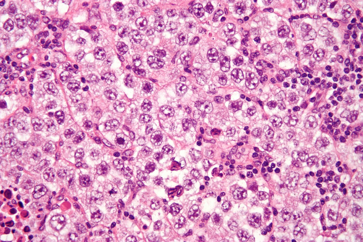
- Histology: Large cells with clear cytoplasm and central nuclei (“Fried egg” appearance).
- Markers: ↑ Placental ALP (pALP); rare ↑ β-hCG. Normal AFP.
- Prognosis/Tx: Excellent prognosis. Highly radiosensitive. Late metastasis.
Mnemonic
Semen eats egg.
Non-Seminomatous Germ Cell Tumors (NSGCT)
- General features: More aggressive, early hematogenous spread, not radiosensitive (chemo/surgery required).
- Yolk Sac (Endodermal Sinus) Tumor
- Epidemiology: Most common testicular tumor in children < 3 years old.
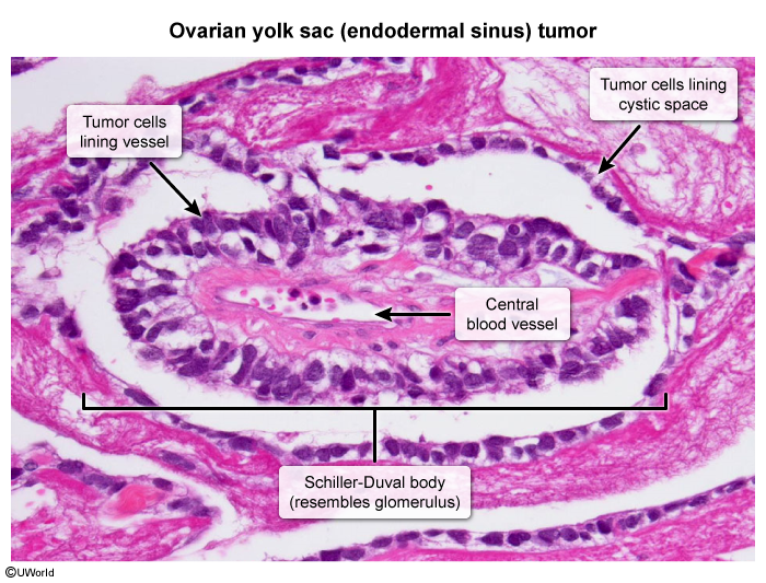
- Histology: Schiller-Duval bodies (resemble primitive glomeruli).
- Markers: ↑ AFP (highly characteristic).
- Choriocarcinoma
- Characteristics: Malignant, disordered proliferation of syncytiotrophoblasts and cytotrophoblasts (placental tissue).
- Spread: Early hematogenous spread to lungs (“Cannonball metastases”) and brain. t
- Markers: ↑↑ β-hCG.
- Clinical: Gynecomastia (hCG mimics LH); Hyperthyroidism (hCG mimics TSH, due to shared α-subunit).
- Embryonal Carcinoma
- Characteristics: Malignant, painful mass with hemorrhage and necrosis.
- Histology: Glandular/papillary morphology.
- Markers: ↑ hCG, ± ↑ AFP (if mixed).
- Teratoma t
- Characteristics: Contains tissue from all 3 germ layers (neural, muscle, cartilage).
- Adults: Generally malignant.
- Children: Generally benign.
Tip
HCG is always elevated in choriocarcinoma and sometimes elevated in seminoma. AFP is always elevated in yolk sac tumors. Both AFP and HCG may be elevated in mixed germ cell tumors.
Non–germ cell tumors
Leydig cell tumor
- Microscopy: Reinke crystals (eosinophilic cytoplasmic inclusions)

Extragonadal germ cell tumors
- Definition: primary germ cell tumors that arise outside of the gonads, anywhere along the body’s midline from the pineal gland to the coccyx.
- Epidemiology: 5–10% of all germ cell tumors; mostly affects young males
- Location
- Midline organs
- Mediastinal > retroperitoneal > intracranial (pineal gland and suprasellar region)
- Sacrococcygeal teratomas
- Most common in infancy or early childhood
- Symptoms
- Chest pain, dyspnea (on exertion), cough
- Diagnosis
- Testicular ultrasound: to rule out gonadal primary tumor
- Tumor markers: alpha fetoprotein (AFP) and human chorionic gonadotropin (HCG)
- Tumor biopsy (confirmatory)