Etiology
- Elderly individuals experience age-related cerebral atrophy. This brain shrinkage puts increased tension on the bridging veins, making them highly susceptible to tearing from shearing forces, even with relatively minor head trauma (like a fall).
- Traumatic SAH: traumatic brain injury
- Nontraumatic (spontaneous) SAH
- Ruptured intracranial aneurysms
- Most commonly occur in the circle of Willis
- Berry aneurysms account for approx. 80% of cases of nontraumatic SAH. c
- Also known as Saccular aneurysm because of the shape
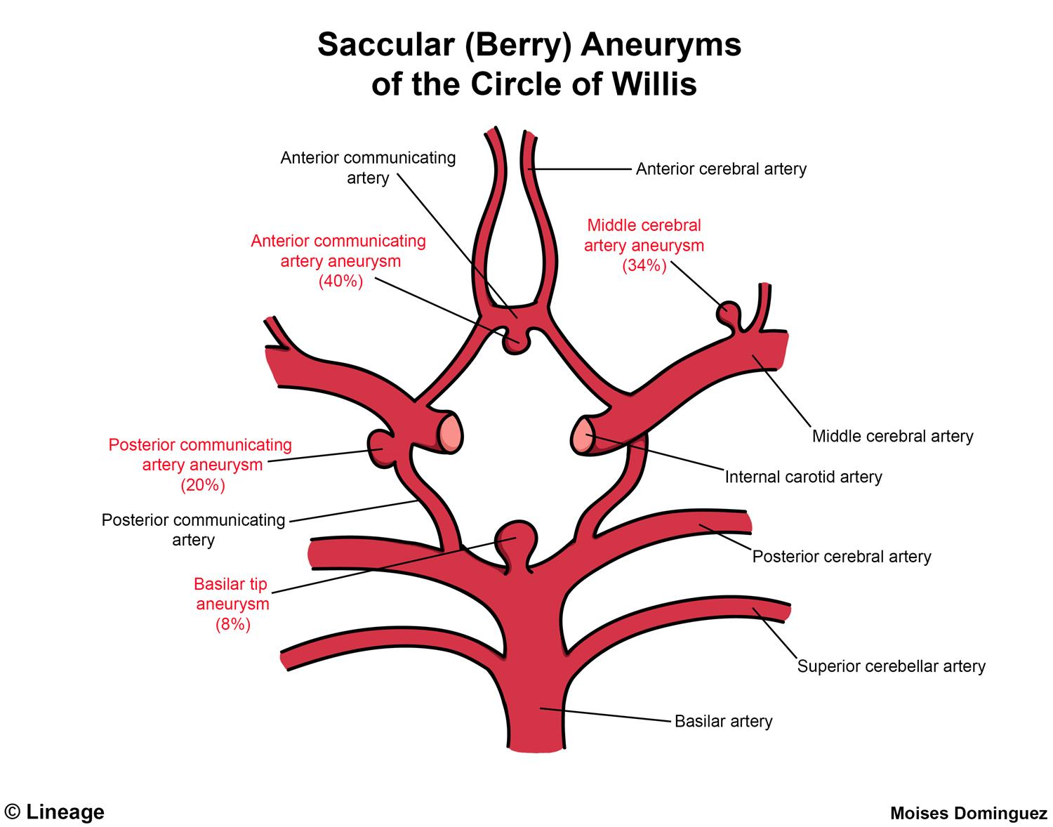
- Round, saccular shape
- Most common type of cerebral aneurysm
- Typically occur at vessel junctions in the circle of Willis, most commonly between the anterior communicating artery and anterior cerebral artery
- Account for ∼ 80% of cases of nontraumatic subarachnoid hemorrhage
- Also known as Saccular aneurysm because of the shape
- Ruptured arteriovenous malformations (AVM)
- Ruptured intracranial aneurysms
Charcot-Bouchard aneurysms vs Saccular (berry) aneurysms
Feature Charcot-Bouchard Saccular (Berry) Etiology Chronic HTN Congenital weakness + Hemodynamics Pathology Lipohyalinosis of microvessels Lacking Internal Elastic Lamina & Media Location Deep Brain (Basal Ganglia, Thalamus) Circle of Willis Bifurcations (ACom > PCom) Vessels Lenticulostriate arteries Medium-sized arteries Rupture Intraparenchymal Hemorrhage Subarachnoid Hemorrhage Symptoms Focal deficits (Hemiparesis) “Thunderclap” Headache, Meningismus Associations Lacunar strokes ADPKD, Ehlers-Danlos, Marfan
-aneurysms/../../../appendix/pasted-image-20250304102040.png)
Link to original
- Saccular: This term means “resembling a sac.” Saccular aneurysms are outpouchings or bulges on one side of a blood vessel wall.
- Berry: The “berry” description refers to the characteristic round shape of these aneurysms. They look like a berry connected to the main artery.
-aneurysms/../../../appendix/pasted-image-20240526113641.png)
Clinical Features
- History:
- “Worst headache of my life” / “Thunderclap” headache (hyperacute onset, reaching maximal intensity within seconds to minutes).
- Transient LOC, nausea, vomiting, syncope, photophobia.
- Sentinel headache: Warning leak occurring days-to-weeks prior in ~10-40% of cases.
- Physical Examination:
- Meningismus (nuchal rigidity, (+) Kernig, (+) Brudzinski signs; can take 6-12 hrs to develop).
- CN III palsy with pupillary involvement (fixed, dilated pupil + “down and out” eye) suggests PCom aneurysm compression.
- Subhyaloid/preretinal hemorrhages (Terson syndrome).
- Focal neurological deficits or depressed GCS (indicates elevated ICP or intraparenchymal extension).
Diagnosis
- Initial / Screening Imaging:
- Non-Contrast Head CT (NCCT): c2
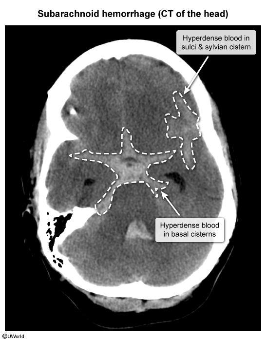
- Sensitivity >95–98% within first 6–12 hrs (drops significantly after 24–48 hrs because CSF dilutes blood).
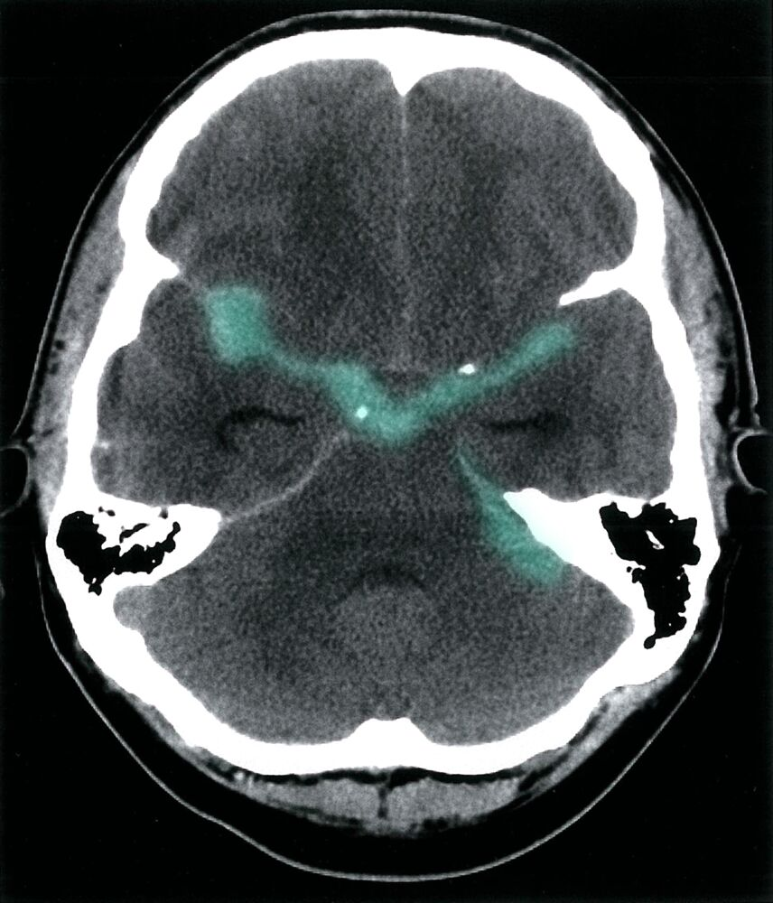
- Hyperattenuation/blood seen in basal cisterns, sulci, and Sylvian fissures.
- Non-Contrast Head CT (NCCT): c2
- Next Step if NCCT is Negative / Equivocal & Suspicion High:
- Lumbar Puncture (LP): c
- Xanthochromia (yellow supernatant from RBC lysis/bilirubin; peaks 6–12 hrs, gold standard LP marker; centrifuge CSF to assess).
- Elevated RBC count that persists from tube 1 to tube 4 (differentiates SAH from traumatic tap).
- Elevated opening pressure (>200 mm H₂O).
- Lumbar Puncture (LP): c
- Confirmatory / Vascular Mapping:
- CT Angiography (CTA): Rapid, non-invasive initial vascular imaging for aneurysm localization.
- Digital Subtraction Angiography (DSA): Gold standard for identifying aneurysm anatomy, source of bleeding, and planning intervention.
- Key Labs:
- Baseline CBC, PT/INR, PTT, Type and Screen.
- BMP (serial monitoring for hyponatremia).
- Cardiac enzymes and ECG (rule out neurogenic stunned myocardium/arrhythmias).
Treatment
Initial management
- Prevention of rebleeding
- Anticoagulant reversal
- Management of blood pressure and cerebral perfusion pressure
- Target SBP < 160 mm Hg
- Other neuroprotective measures
- Start ICP management (e.g., elevate head 30°, IV mannitol, short-term controlled hyperventilation).
Treatment of aneurysmal SAH
- Intracranial aneurysm repair
- Endovascular coiling
- Microsurgical clipping
- Prevention of vasospasm and delayed cerebral ischemia
- Administer oral nimodipine
- Only administer nimodipine orally or via enteral tube; Parenteral administration is associated with significant adverse effects (e.g., severe hypotension and cardiac arrest).
- Treatment of hydrocephalus: may include an external ventricular drain (EVD), lumbar drainage, or permanent ventriculoperitoneal shunt
- Administer oral nimodipine
Warning
Generally avoid nitrates for blood pressure control in brain injury, as they may elevate ICP. Consider alternative agents (e.g., titratable nicardipine or labetalol).
Complications
Vasospasm
- Occurs in approx. 30% of patients with SAH
- Pathophysiology
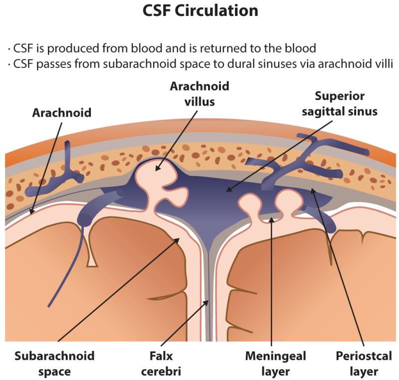
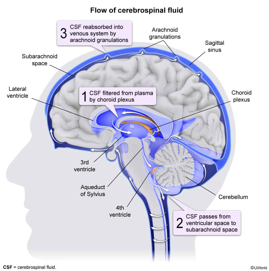
- Impaired CSF reabsorption from the arachnoid villi → nonobstructive (communicating) hydrocephalus → ↑ intracranial pressure → ↓ cerebral perfusion pressure → ischemia
- Release of clotting factors and vasoactive substances → diffuse vasospasm of cerebral vessels → ischemia
- Impaired CSF reabsorption from the arachnoid villi → nonobstructive (communicating) hydrocephalus → ↑ intracranial pressure → ↓ cerebral perfusion pressure → ischemia
- Can lead to ischemic stroke
- Most common in patients with nontraumatic SAH due to a ruptured aneurysm
- Usually occurs between 3–10 days after SAH
- Prevention & Treatment
- Nimodipine (Calcium Channel Blocker): t
- Standard of Care: Started on admission (Day 1) for all SAH patients.
- Mechanism: It blocks this calcium influx into the neurons, preventing cellular death. Prevents ischemic neurological deficits and improves mortality (neuroprotection).
- Note: It does not necessarily prevent the angiographic vasospasm itself, but prevents the cellular injury associated with it.
- Hemodynamic Augmentation (formerly “Triple H”):
- Current Goal: Maintain Euvolemia and Induced Hypertension (using vasopressors like phenylephrine or norepinephrine).
- Rationale: ↑ MAP pushes blood through the narrowed vessels to maintain Cerebral Perfusion Pressure (CPP).
- Refractory Cases:
- Intra-arterial vasodilators (e.g., verapamil, nicardipine).
- Balloon angioplasty (mechanical dilation).
- Nimodipine (Calcium Channel Blocker): t