Definition: head trauma through strong rotational and shearing force
Epidemiology
high mortality and a significant cause of death
Etiology
violent shaking of a child
Pathophysiology
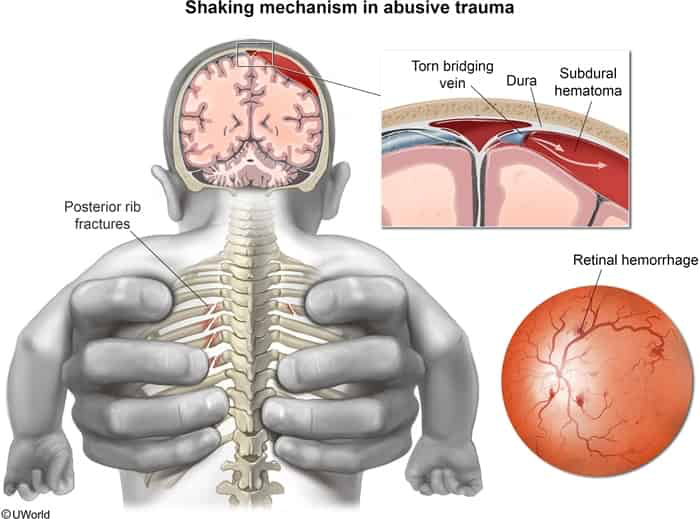
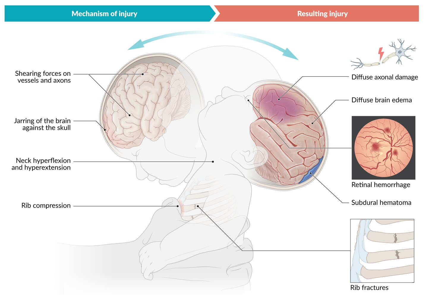
- Rotational and shearing forces → shearing off of bridging veins → subdural hematoma
- Shaking of the child with a weak neck support → respiratory problems and apnea → hypoxia → brain edema and ischemia → diffuse axonal damage
Clinical features
- Classic Triad:
- Subdural hematoma (SDH) (especially interhemispheric or convexities).
- Retinal hemorrhages (bilateral, multi-layered, extending to periphery/ora serrata). c
- Encephalopathy (lethargy, irritability, altered mental status, coma).
- History Clues:
- Plausible injury mechanism absent or inconsistent with developmental stage (e.g., “rolled off a low bed/couch” causing severe intracranial hemorrhage).
- Discrepant, evolving, or delayed history provided by caregivers.
- Physical Examination:
- Neurological: Bulging/tense anterior fontanelle, rapidly increasing head circumference, altered mental status (AMS), seizures, apnea/hypopnea, posturing (decorticate/decerebrate).
- Ophthalmologic: Extensive, multi-layered retinal hemorrhages, retinoschisis, retinal detachment.
- Cutaneous: Bruising in non-mobile infants (“TEN-4 Rule”: Torso, Ears, Neck in children < 4 mo) or patterned bruises.
- Skeletal:
- Metaphyseal corner / “bucket-handle” fractures (pathognomonic; from pulling/twisting forces).
- Posterior rib fractures (pathognomonic; from thoracic squeezing during shaking).
- Fractures of varying ages/healing stages; skull fractures (complex, diastatic, crossing sutures).
Diagnostics
- Non-contrast CT
- Subdural hematomas and/or subarachnoid hemorrhage of varying ages
- Reversal sign: diffuse blurring of the grey-white matter interface
- Diffuse punctate hemorrhages
Differential Diagnostics
- Accidental Head Trauma:
- Diff by history consistent with developmental ability; falls < 3–4 ft typically cause simple, linear parietal skull fractures without complex SDH, DAI, or severe bilateral multi-layered retinal hemorrhages. c
- Osteogenesis Imperfecta (OI):
- Diff by blue sclerae, dentinogenesis imperfecta, positive family history, wormian bones on skull radiographs; does not cause subdural hemorrhage or retinal hemorrhages.
- Congenital Bleeding Disorders (e.g., Hemophilia, vWD, Vit K Deficiency):
- Diff by abnormal coagulation cascade labs (↑ PTT or ↑ PT) and absence of specific NAT-associated fracture patterns (e.g., posterior rib/bucket-handle fractures).
- Glutaric Aciduria Type 1:
- Diff by macrocephaly, frontotemporal atrophy (“bat wing” appearance on CT/MRI), basal ganglia signal abnormalities, and elevated urine glutaric acid on organic acid screening.
- Benign Enlargement of Subarachnoid Spaces (BESS):
- Diff by macrocephaly with prominent frontal subarachnoid space, normal brain parenchyma, preserved neurodevelopment, and absence of retinal hemorrhages.
Treatment
- Always notify Child Protective Services.
- Medical practitioners are obligated to report child abuse even if that means breaking patient-physician confidentiality.
- Reasonable suspicion is sufficient; confirmation of abuse or neglect is not required for reporting purposes.
- Interview child and parent/caregiver separately if possible.
- Keep verbatim record.