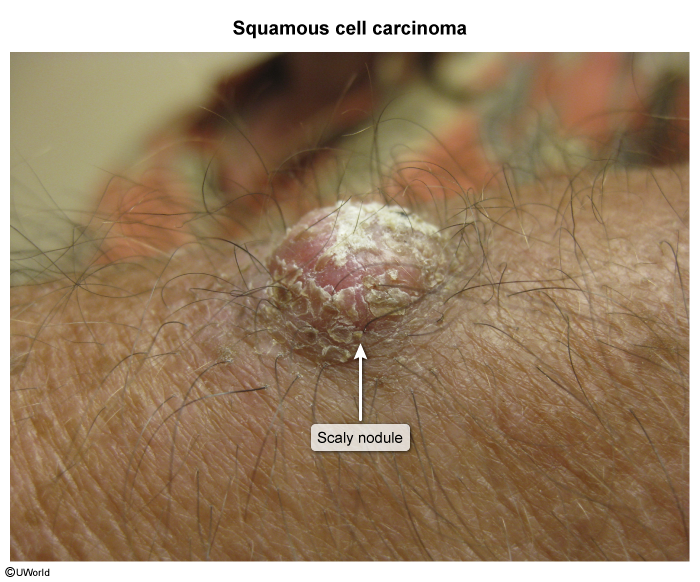
Epidemiology
Etiology
- Cumulative UV exposure (from the sun or from tanning beds) is the most significant risk factor
Pathophysiology
Clinical features
- History: Rapidly enlarging, non-healing lesion that frequently bleeds, scabs, or becomes painful; typically located on sun-exposed sites (head, neck, lower lip, ears, dorsum of hands).
- Physical Exam:
- Classic lesion: Erythematous, indurated, scaly plaque or firm nodule with central crusting, hyperkeratosis, or ulceration.
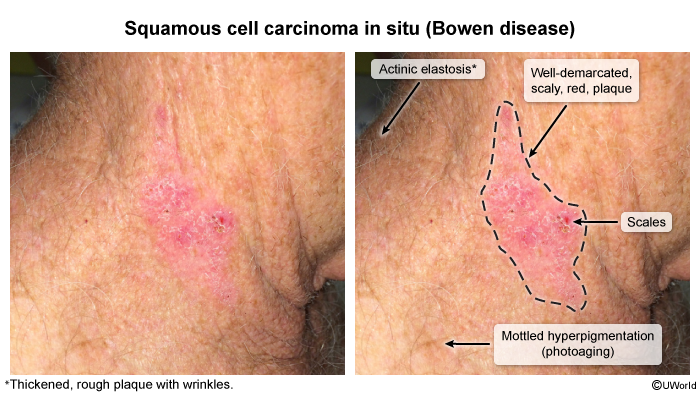
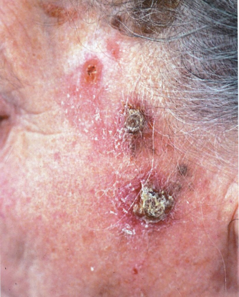
- Cutaneous horn: Projection of cohesive keratin overlying the tumor base.
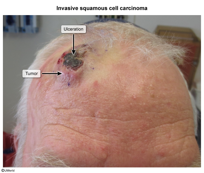
- Keratoacanthoma variant: Rapidly growing (over weeks), dome-shaped, flesh-colored to red nodule with a central keratin-filled plug/crater (often spontaneously regresses, but treated as well-differentiated cSCC).
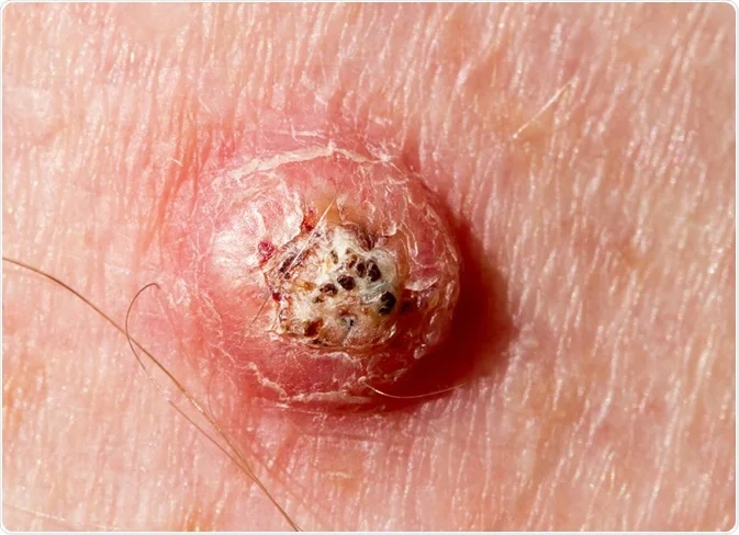
- Perineural invasion: Numbness, paresthesias, localized pain, or motor nerve palsy (indicates advanced/high-risk disease).
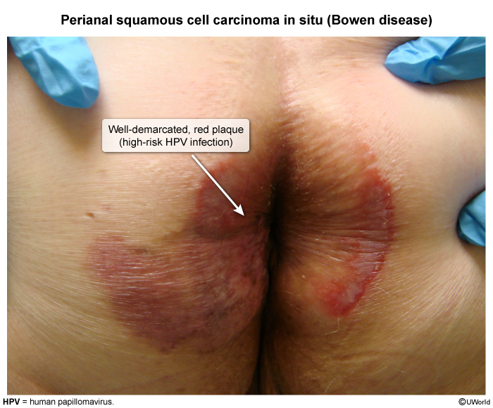
- Classic lesion: Erythematous, indurated, scaly plaque or firm nodule with central crusting, hyperkeratosis, or ulceration.
Subtypes and variants
Marjolin ulcer
- Etiology & Risk Factors:
- Malignant transformation of chronic non-healing wounds or scars.
- Most common: Burn scars (classic); also chronic osteomyelitis sinus tracts and venous ulcers. c
- Latency: Long interval (10–30+ years post-initial injury).
- Clinical Presentation:
- Chronic wound with sudden changes: rapid growth, increased pain, or foul discharge.
- PE: Everted/rolled borders, friable tissue, bleeding on contact, or an exophytic/fungating mass.
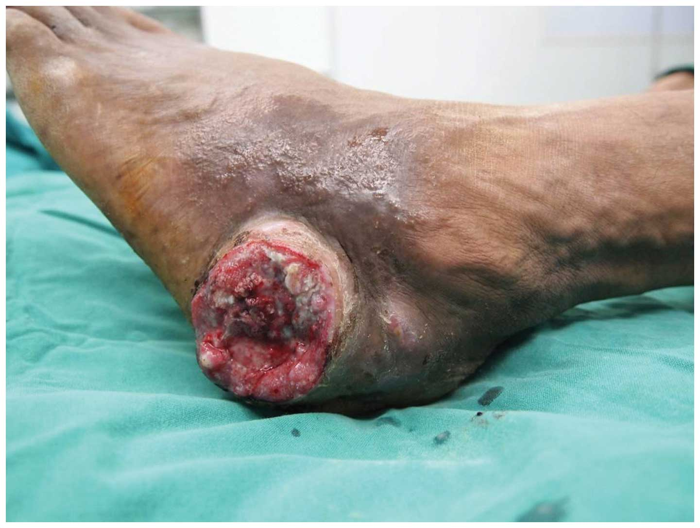
- Diagnosis:
- Gold Standard: Biopsy of the ulcer edge/margin (punch or incisional).
- Histology: Aggressive Squamous Cell Carcinoma (SCC).
- Imaging: MRI to assess deep soft tissue/bone invasion; CT staging for regional LN or distant metastases.
- Key Differentials:
- Venous stasis ulcer: Medial malleolus, hemosiderin deposition, flat borders (non-everted).
- Pyoderma gangrenosum: Rapidly progressive, violaceous undermined edges, pathergy (+), sterile neutrophilic infiltrate on bx.
- Management:
- First-line: Wide local excision (WLE) with ≥1–2 cm margins +/- regional LN assessment.
- Advanced/Invasive: Amputation if bone, joint, or major neurovascular bundle involved.
- Adjuvant: Radiotherapy for close/positive margins, perineural invasion, or nodal disease.
- High-Yield Exam Pearl:
- Carries a significantly higher rate of metastasis (~30%) and recurrence than conventional cutaneous SCC.
Diagnostics
Pathology
cSCC occurs as a result of malignant transformation of keratinocytes in the stratum spinosum (prickle cell layer) of the epidermis. These atypical keratinocytes appear as enlarged, polygonal cells with nuclear pleomorphism and atypical mitoses.
- Sheets or nests of malignant keratinocytes
- Keratinization (eg, keratin pearls) & intercellular bridges
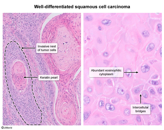
- Keratin pearls: An abnormal structure formed by concentric layers of epithelial cells surrounding a keratin matrix secreted by the cells. Associated with well-differentiated squamous cell carcinoma.
Differential diagnostics
| Feature | Basal Cell Carcinoma (BCC) | Cutaneous Squamous Cell Carcinoma (cSCC) |
|---|---|---|
| Origin | Basal cells in the epidermis | Squamous cells in the epidermis |
| Common Locations | Sun-exposed areas: face, neck | Sun-exposed areas: face, ears, neck, lips, back of the hands |
| Appearance | Pearly or waxy bump, flat flesh-colored or brown scar-like lesion | Firm, red nodule, flat lesion with a scaly, crusted surface |
| Growth Rate | Slow-growing | Can be faster-growing than BCC |
| Risk of Metastasis | Rare, low risk | Higher risk compared to BCC |
| Common Causes | Chronic sun exposure, fair skin | Chronic sun exposure, fair skin, history of burns or radiation exposure |
| Typical Age Group | Older adults | Older adults |
| Prevalence | Most common type of skin cancer | Second most common type of skin cancer |
| Treatment Options | Surgical removal, topical medications, radiation therapy | Surgical removal, topical medications, radiation therapy, chemotherapy for advanced cases |
| Prognosis | Generally good if detected early | Good if detected early; can be aggressive if untreated |
- Nodular BCC classically has a translucent or pearly appearance, often with central telangiectasias.
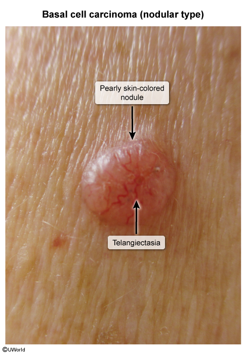
- Cutaneous SCC classically has a rough or scaly (keratinized) surface