Epidemiology
Etiology
Pathophysiology
Clinical features
- History:
- Preceding viral prodrome (fever, malaise, myalgias, URI or GI symptoms days to weeks prior).
- Acute or subacute HF symptoms: Dyspnea on exertion, orthopnea, PND, fatigue.
- Chest pain (often sharp, pleuritic if concurrent perimyocarditis; or ischemic-like).
- Palpitations, presyncope, or syncope (secondary to arrhythmias).
- Physical Exam:
- Sinus tachycardia out of proportion to fever or clinical state.
- Signs of HF: Elevated JVP, S3/S4 gallop, bibasilar crackles, peripheral edema, hepatomegaly.
- Holosystolic murmur at apex (functional mitral regurgitation due to LV dilation).
- Pericardial friction rub (if perimyocarditis).
- Cardiogenic shock: Cold extremities, hypotension, weak peripheral pulses (pulsus alternans).
Diagnostics
- Initial:
- ECG: Sinus tachycardia, arrhythmias, nonspecific ST-T changes. (Diffuse ST-elevation/PR depression if myopericarditis).
- CXR: Often normal; may show cardiomegaly, pulmonary edema, or pleural effusions.
- Key Labs: ↑ Troponin/CK-MB (indicates myocyte necrosis), ↑ ESR/CRP, ↑ BNP/NT-proBNP.
- Echocardiogram: Shows chamber enlargement, global hypokinesis, ↓ LVEF, or pericardial effusion. Helps rule out structural/valvular disease.
- Confirmatory: Cardiac MRI (Best non-invasive test). Shows late gadolinium enhancement (LGE) indicating edema and myocardial scar.
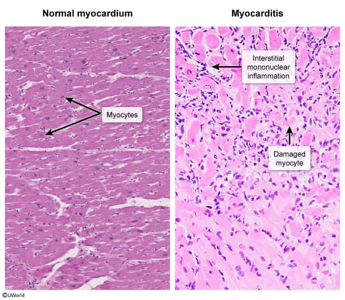
- Gold Standard: Endomyocardial Biopsy (Rarely done). Shows lymphocytic infiltrate w/ myocyte necrosis. Reserved for acute fulminant HF (to r/o Giant cell myocarditis which requires immunosuppression) or failure to respond to standard therapy.
Treatment
Complications