A key differentiator is that upper airway obstruction typically causes stridor (an inspiratory sound), while lower airway disease is characterized by wheezing (an expiratory sound). t
Upper Airway Diseases
The primary sign of upper airway obstruction is stridor, a high-pitched sound caused by turbulent airflow through a narrowed larynx or trachea.
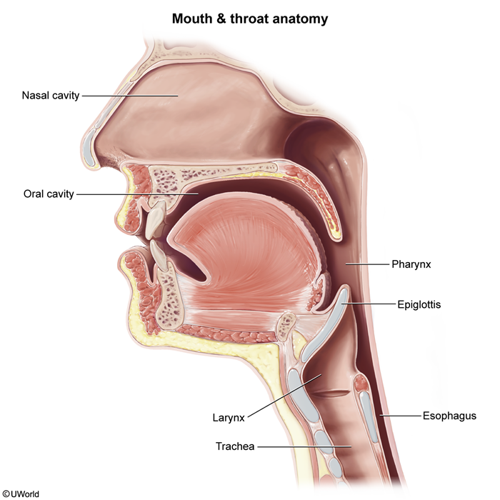
- Croup (Laryngotracheobronchitis)
- Site: Larynx, trachea (subglottic region).
- Etiology: Parainfluenza virus.
- Presentation: Age 6mo-3yr. Barking seal-like cough, inspiratory stridor.
- Dx: Clinical. X-ray: Steeple sign.
- Tx: Corticosteroids (dexamethasone). Nebulized epinephrine if severe.
- Epiglottitis
- Site: Epiglottis and supraglottic structures.
- Etiology: Hib (less common post-vaccine).
- Presentation: Age 2-7yr. Acute high fever, drooling, tripod position. Medical emergency.
- Dx: Lateral neck X-ray: Thumbprint sign. Do NOT examine throat.
- Tx: Secure airway in OR. Ceftriaxone.
- Foreign Body Aspiration
- Site: Right mainstem bronchus is most common. Can be anywhere in tracheobronchial tree.
- Presentation: Toddler (1-3yr). Sudden choking. Unilateral wheezing or diminished breath sounds.
- Dx/Tx: Rigid bronchoscopy.
Lower Airway Diseases
Lower airway diseases involve the bronchi and bronchioles, typically presenting with wheezing, tachypnea, and increased work of breathing.
- Bronchiolitis
- Site: Small airways (bronchioles).
- Etiology: RSV.
- Presentation: <2 yr old. URI sx followed by tachypnea, diffuse wheezing/crackles. t
- Tx: Supportive: nasal suctioning, O2, hydration.
- Prophylaxis: Palivizumab for high-risk infants.
- Asthma
- Site: Bronchi (reversible bronchoconstriction).
- Presentation: Recurrent wheezing, cough (worse at night).
- Tx: Acute: Albuterol (SABA). Chronic: Inhaled corticosteroids (ICS).
Parenchymal Disease
- Pneumonia
- Site: Lung parenchyma (alveoli).
- Etiology: Viral (RSV) or bacterial (S. pneumoniae).
- Presentation: Fever, cough, tachypnea. Exam shows crackles or focal decreased breath sounds.
- Tx: Viral is supportive. Bacterial is Amoxicillin.
- ARDS
- Site: Alveolar-capillary membrane.
- Etiology: Sepsis, pneumonia → diffuse alveolar damage.
- Presentation: Severe, refractory hypoxemia (PaO2/FiO2 < 300).
- Dx: CXR: Bilateral pulmonary infiltrates (“white-out”).
- Tx: Low tidal volume ventilation + PEEP.