- Contents
- Males: Spermatic cord.
- Females: Round ligament of the uterus.
- Both:
- Ilioinguinal nerve (L1): Enters partway through the canal and exits via the superficial ring. Does NOT pass through the deep ring. Provides sensation to the upper medial thigh, root of the penis/scrotum, and mons pubis/labia majora. Most commonly injured nerve in hernia repair.
- Genital branch of the genitofemoral nerve (L1, L2): Supplies the cremaster muscle and scrotal skin (males) or labia majora (females).
Etiology
Pathophysiology
Indirect Inguinal Hernia
- Pathophysiology:
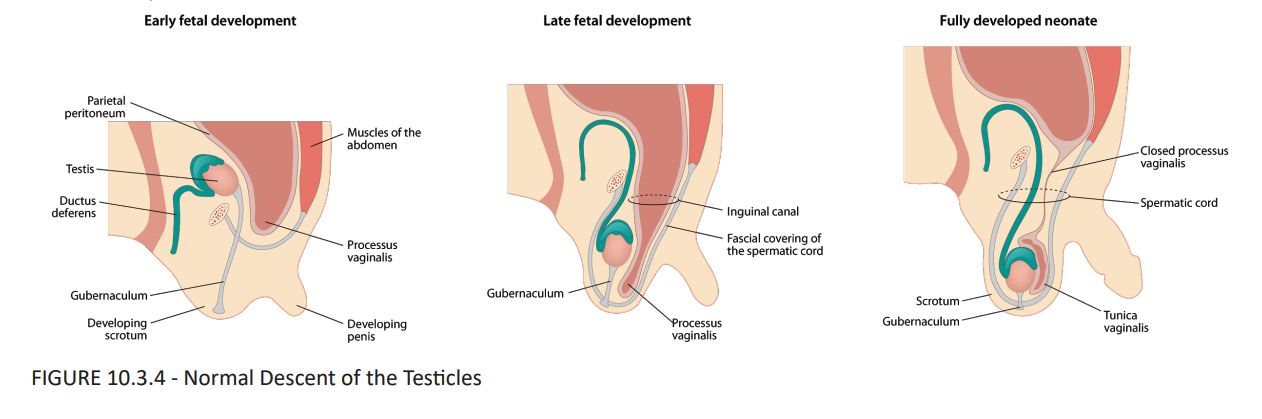
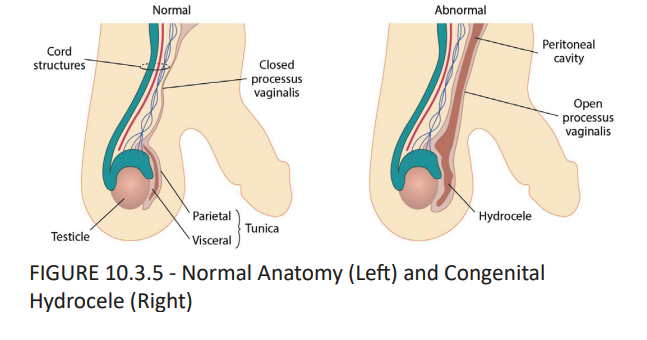
- Results from a patent processus vaginalis, which is a congenital failure of the peritoneal outpouching to close.
- The herniating bowel follows the path of the spermatic cord through the inguinal canal.
- Course:
- Enters the deep inguinal ring, traverses the entire inguinal canal, and exits through the superficial inguinal ring. It can descend into the scrotum.
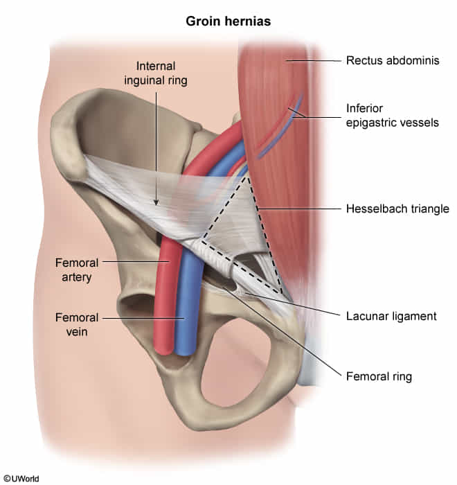
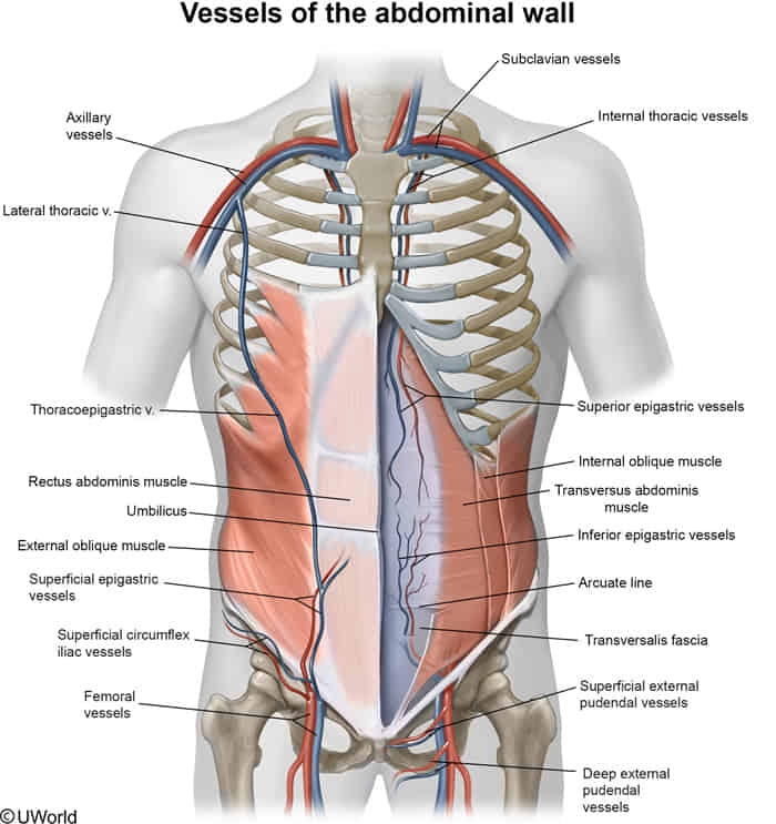
- Always located LATERAL to the inferior epigastric vessels.
- Epidemiology:
- The most common type of inguinal hernia (about 80%).
- More common in children and young males.
- Associations:
- Covered by all three layers of spermatic fascia (internal spermatic, cremasteric, and external spermatic).
Direct Inguinal Hernia
- Pathophysiology:
- Protrudes through a weakness in the posterior wall of the inguinal canal (transversalis fascia).
- This area of weakness is known as Hesselbach’s triangle.
- Typically an acquired condition due to mechanical stress or weakness of the abdominal wall, common in older men.
- Hesselbach’s Triangle Borders:
- Medial: Lateral border of the rectus abdominis muscle.
- Superolateral: Inferior epigastric vessels.
- Inferior: Inguinal ligament.
- Course:
- Pushes directly through the floor of the canal and exits through the superficial inguinal ring. It does not traverse the deep ring.
- Always located MEDIAL to the inferior epigastric vessels.
- Rarely enters the scrotum.
Clinical features
- History: c
- Groin bulge or heaviness, typically exacerbated by standing, coughing, or Valsalva maneuver; improves or disappears when supine.
- Aching groin discomfort; sharp severe pain suggests acute complication.
Differential diagnostics
Rectus sheath hematoma
- Etiology & Risk Factors:
- Rupture of inferior epigastric artery (below the arcuate line) or superior epigastric artery.
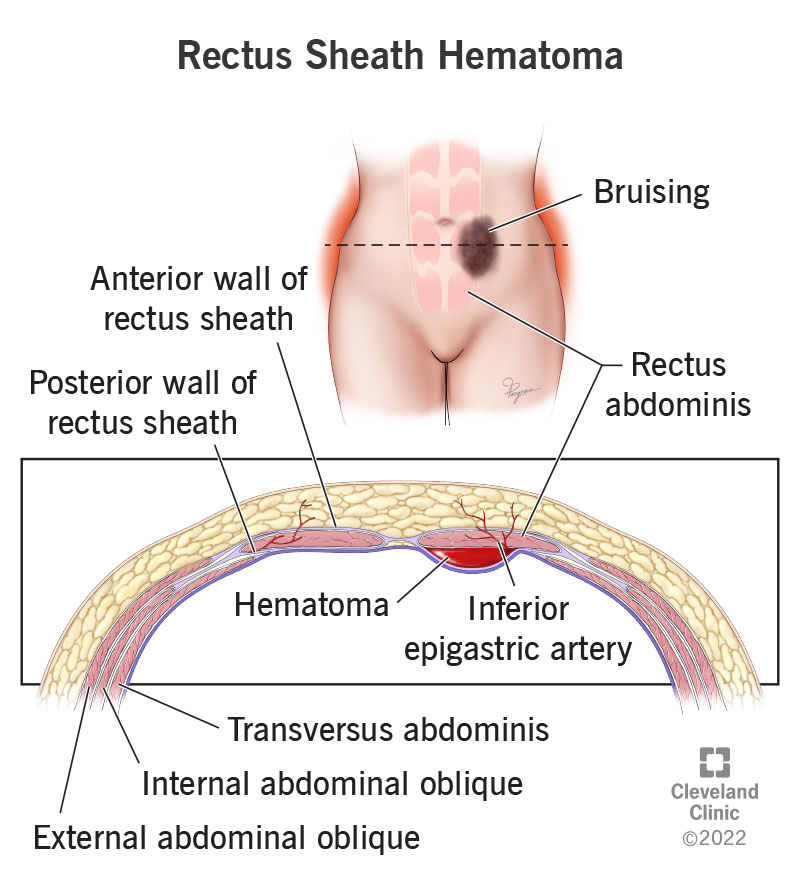
- Risk factors: Anticoagulation/antiplatelet use + sudden ↑ intra-abdominal pressure (severe coughing, vomiting, straining, trauma). c
- Rupture of inferior epigastric artery (below the arcuate line) or superior epigastric artery.
- Clinical Presentation:
- Acute, sharp, well-localized, unilateral abdominal pain with a tender, palpable abdominal wall mass.
- Fothergill sign (+): Mass remains palpable and fixed, does not cross midline when pt flexes rectus muscles (e.g., neck/leg raise).
- Carnett sign (+): Tenderness persists or worsens with abdominal muscle contraction (rules out visceral pain).
- Severe cases: Tachycardia, hypotension, syncope (hypovolemic shock).
- Diagnosis:
- Best/Confirmatory: CT Abdomen & Pelvis with IV contrast (detects hematoma, excludes intra-abdominal pathology, identifies active contrast “blush”).
- Bedside/Initial (if unstable): Abdominal Ultrasound.
- Labs: Serial ↓ Hb/Hct, ↑ PT/INR, ↑ PTT, type & screen.
- Differentiating Features:
- Appendicitis/Diverticulitis: Fothergill (-), Carnett (-), systemic signs (fever, altered bowel habits), CT shows bowel inflammation.
- Incarcerated Hernia: Palpable fascial defect with herniated bowel/omentum on CT.
- Management Hierarchy:
- Hemodynamically Stable / Non-expanding:
- Conservative: Rest, analgesia, serial CBC monitoring.
- Hold and reverse anticoagulation (e.g., 4F-PCC + IV Vitamin K for warfarin; Protamine for heparin).
- Hemodynamically Unstable / Active Bleed / Expanding:
- IVF resuscitation, pRBC/blood product transfusion.
- Transcatheter Angiographic Embolization (TAE) (treatment of choice for active extravasation).
- Refractory / Surgical Indications:
- Surgical ligation/evacuation: Only if embolization fails or pt develops Abdominal Compartment Syndrome.
- Hemodynamically Stable / Non-expanding:
Diagnostics
Treatment
Complications
Postoperative complications
- Inguinodynia; paresthesia or anesthesia over the surgical site
- Can be secondary to nerve injury during the surgical procedure (i.e., transection of the ilioinguinal nerve or genitofemoral nerve that traverse the inguinal region) or due to nerve entrapment within the mesh.
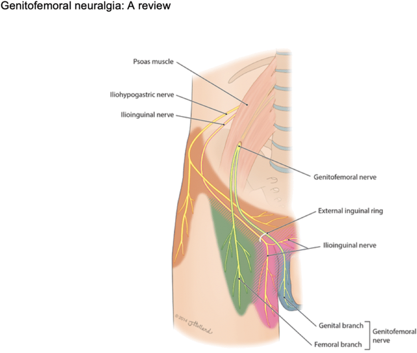
- Genitofemoral nerve
- Can be secondary to nerve injury during the surgical procedure (i.e., transection of the ilioinguinal nerve or genitofemoral nerve that traverse the inguinal region) or due to nerve entrapment within the mesh.